. 2020 Feb 11;17(1):15.
doi: 10.1186/s12987-020-0176-z.

Aquaporin 1 and the Na+/K+/2Cl- cotransporter 1 are present in the leptomeningeal vasculature of the adult rodent central nervous system

Affiliations

Aquaporin 1 and the Na+/K+/2Cl- cotransporter 1 are present in the leptomeningeal vasculature of the adult rodent central nervous system

Qianliang Li et al. Fluids Barriers CNS. .

Abstract

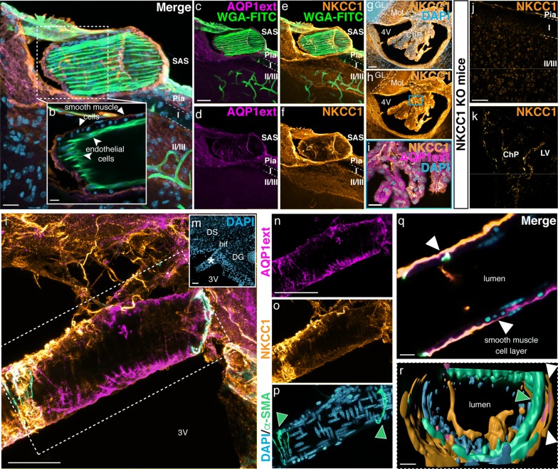
Background: The classical view of cerebrospinal fluid (CSF) production posits the choroid plexus as its major source. Although previous studies indicate that part of CSF production occurs in the subarachnoid space (SAS), the mechanisms underlying extra-choroidal CSF production remain elusive. We here investigated the distributions of aquaporin 1 (AQP1) and Na+/K+/2Cl- cotransporter 1 (NKCC1), key proteins for choroidal CSF production, in the adult rodent brain and spinal cord.

Methods: We have accessed AQP1 distribution in the intact brain using uDISCO tissue clearing technique and by Western blot. AQP1 and NKCC1 cellular localization were accessed by immunohistochemistry in brain and spinal cord obtained from adult rodents. Imaging was performed using light-sheet, confocal and bright field light microscopy.

Results: We determined that AQP1 is widely distributed in the leptomeningeal vasculature of the intact brain and that its glycosylated isoform is the most prominent in different brain regions. Moreover, AQP1 and NKCC1 show specific distributions in the smooth muscle cell layer of penetrating arterioles and veins in the brain and spinal cord, and in the endothelia of capillaries and venules, restricted to the SAS vasculature.

Conclusions: Our results shed light on the molecular framework that may underlie extra-choroidal CSF production and we propose that AQP1 and NKCC1 within the leptomeningeal vasculature, specifically at the capillary level, are poised to play a role in CSF production throughout the central nervous system.

Keywords: Aquaporin 1; Capillaries; Leptomeningeal vasculature; NKCC1; Penetrating arterioles; Subarachnoid space; Veins; Venules.


Conflict of interest statement

The authors declare that they have no competing interests.

Figures

Fig. 1
Fig. 1
uDISCO clearance of the intact mouse head depicts the expression of aquaporin 1. a Mouse brain (P60) cleared by uDISCO and immunolabeled for AQP1 (AQP1int, green) reveals the vasculature network in the leptomeninges, including the middle cerebral arteries (MCA, arrows). AQP1+ cells also line the subarachnoid cisterns and the olfactory bulb. b Optical section reveals AQP1+ choroidal epithelial cells and olfactory ensheathing glia cells. c, d Higher magnification images of the areas depicted in b (blue and purple squares) showing AQP1 in the glomerular layer (arrow) and in choroidal epithelial cells (asterisk). e Representative micrograph of a parasagittal section of an adult mouse brain (P90) immunolabeled for AQP1 (AQP1ext, grey). AQP1ext+ epithelial cells of the choroid plexus are observed in the fourth (f) and in the lateral ventricles (g). In contrast, olfactory ensheathing glia cells in the olfactory bulb are not immunolabeled (h). i Representative micrograph of a coronal section from adult mouse brain (P90) immunolabeled with AQP1 (AQP1int, grey). j Higher magnification of the depicted area in i (square) shows in detail AQP1int+ epithelial cells in the choroid plexus of the lateral ventricles. k Olfactory ensheathing glia cells are also immunoreactive. Dashed line in k depicts the mitral cell layer. l Immunoblotting reveals a band of 35 kDa, corresponding to the glycosylated form of AQP1, detected in the BS, Cb, Ctx, Hip, Hyp and OB, obtained from young adult mice (P30). The non-glycosylated form of AQP1, corresponding to a band of 28 kDa, is detected in choroid plexi and kidney homogenates obtained from young adult mice (P30). The housekeeping protein GAPDH (37 kDa) was used as loading control. Control antigen confirms antibody-epitope specific binding. m Graphic shows the relative AQP1 protein levels, in relation to GAPDH. BS, brain stem; Cb, cerebellum; ChP, choroid plexus; Ctx, cerebral cortex; CPu, caudate putamen; EPL, external plexiform layer; Fi, fimbria; GAPDH, glyceraldehyde 3-phosphate dehydrogenase; GL, glomerular layer; Hip, hippocampus; Hyp, hypothalamus; IC, internal capsule; IPL, internal plexiform layer; Kdy, kidney; LV, lateral ventricle; OB, olfactory bulb; PirCtx, piriform cortex, SCh, suprachiasmatic nuclei; Thal, thalamus; WM, white matter; 3V, third ventricle; 4V, fourth ventricle. Scale bars: a, b, e 1 mm; c, i 500 μm; d, 200 μm; fh, j, k 50 μm
Fig. 2
Fig. 2
AQP1 is expressed in the brain and peripheral vasculature. a Confocal micrograph from an adult mouse brain (P90) immunolabeled for AQP1 (AQP1ext, magenta and AQP1int, green). DAPI nuclear counterstaining (blue). b AQP1ext+ blood vessel, located around the ventricles (delimited by the magenta square in a). cf Immunoreactive epithelial choroid plexus cells, located in the lateral ventricles, are labeled with both antibodies (high magnification of the area delimited by the green square in a). g, h Micrographs of mouse kidney show the distribution of AQP1 in the vascular endothelium and proximal tubules. i, j Higher magnification image of a blood vessel immunolabeled for CD31 (green) and AQP1int (magenta) (delimited by square in h). Asterisk indicates the lumen of a blood vessel and arrows indicate proximal tubules. k, l AQP1+ endothelial cells are also detected in the heart of adult mice. mo Paraffin sections obtained from adult rat brain show AQP1 immunoreactive blood vessels in the hippocampal fissure and epithelial cells of the choroid plexus located in the third ventricle. Arrows and curved arrowheads indicate arterioles or veins and capillaries or venules, respectively. Straight arrowheads indicate AQP1 blood vessels. 3V, third ventricle; BV, blood vessel; ChP, choroid plexus; DG, dentate gyrus; LV, lateral ventricle; PT, proximal tubule. Scale bars: a, b and gj 50 µm; cf 5 µm; k 1 mm; l 100 µm; m 2 mm; n 500 μm; o 200 μm
Fig. 3
Fig. 3
AQP1 and NKCC1 are expressed by the choroidal epithelial cells and in the leptomeningeal vasculature. af Confocal micrograph show a leptomeningeal WGA-FITC+ (green) labeled vessel immunoreactive for AQP1 (magenta) and NKCC1 (orange) in the adult mouse brain (P90). In b an optical section reveals that AQP1+/NKCC1+ cells are restricted to the smooth muscle cell layer (arrowheads) and absent in the endothelial cells (curved arrowheads), which are labeled by WGA-FITC. g, h NKCC1 is detected in the choroid plexus epithelia, in ependymal cells and in the molecular layer of the cerebellum, as shown in the micrographs of the fourth ventricle. i Double labeling confirms AQP1 and NKCC1 presence in choroid plexus epithelial cells (higher magnification of the area delimited by the blue square in h). j, k Brain sections obtained from NKCC1 KO adult mice show no immunoreactivity in the brain parenchyma neither in the choroid plexus. l, m Histological sections immunolabeled with antibodies against AQP1ext (magenta), NKCC1 (yellow) and α-SMA (cyan), reveal AQP1ext+/NKCC1+/α-SMA+ leptomeningeal vessels around the hippocampus and third ventricle. Low magnification micrograph shows DAPI (blue) counterstaining and indicates a leptomeningeal blood vessel (asterisk) closely located to the hippocampal fissure. np Higher magnification of an AQP1ext+/NKCC1+ vessel (delimited by the dashed square in j. Arrowheads indicate α-SMA+ cells. (q) Optical sectioning reveals that both AQP1 and NKCC1 are distributed in the smooth muscle cell layer (arrowheads). r 3D rendering of the leptomeningeal vessel confirms AQP1 and NKCC1 restriction to the smooth muscle cell layer (arrowheads). ChP, choroid plexus; DG, dentate gyrus; DS, dorsal subiculum; GL, granular layer; hif, hippocampal fissure; Mol, molecular layer; SAS, subarachnoid space; 3V, third ventricle, 4V, fourth ventricle. Scale bars: a, i 20 µm; bf, q, r 10 µm; g, h, jp 50 µm
Fig. 4
Fig. 4
AQP1 and NKCC1 are present in smooth muscle and endothelial cells of the leptomeningeal vasculature. a, b Paraffin sections of adult mouse brain (P90) immunolabeled with anti-AQP1int or anti-NKCC1 (both brown). c Some sections were stained with hematoxylin (HE, pink) and the vascular identity of blood vessels located in the subarachnoid space (cisterna interpendicularis, delimited by square in a, b) was determined. d, e Consecutive sections show that AQP1int+/NKCC1+ cells are present in the smooth muscle cell layer of arterioles (arrowheads) and in the endothelium of capillaries and venules, respectively (curved arrowheads). f, g Vascular endothelial cells were labeled by lectin (WGA-FITC, green), followed by standard Immunolabeling. DAPI counterstain (blue) reveal the location of the leptomeningeal vessel (asterisk). hj Higher magnification confocal images show that AQP1 is restricted to tunica media, where AQP1ext+ smooth muscle cells, identified by their round soma (arrowheads) are observed, whereas AQP1 is not present in the endothelial cell layer (curved arrowheads). The arrow indicates a leptomeningeal cell, also AQP1ext+. BS, brain stem; Cb, cerebellum; cp, cerebral peduncle; Ctx, cerebral cortex; Hip, hippocampus; Hyp, hypothalamus; OB, olfactory bulb; Pn, pontine nuclei. Scale bars: a, b 2 mm; ce 100 μm; fj 50 μm
Fig. 5
Fig. 5
AQP1 and NKCC1 are present in leptomeningeal vascular endothelia of the spinal cord. Micrographs of paraffin sections obtained from the spinal cord of adult mice (P90) and immunolabeled for AQP1 and NKCC1 (brown). AQP1 immunoreactivity is predominantly located in C fibers in the dorsal horns of the spinal cord (a, arrowheads), whereas NKCC1 is observed throughout the spinal cord grey matter (d). b, e High magnification of the area delimited by the blue rectangle in a and d, respectively, show AQP1int+/NKCC1+ leptomeningeal vessels (arrows) in the spinal cord. c, f High magnification micrographs of the area delimited by the green squares in b and e show AQP1int+/NKCC1+ cells in the vascular endothelium, restricted to the subarachnoid space along the spinal cord (curved arrowheads). DRG, dorsal root ganglia; SAS, subarachnoid space. Scale bars: a, d 1 mm; b, e 100 μm; c, f 50 μm
Fig. 6
Fig. 6
AQP1 and NKCC1 distribution in the CNS leptomeningeal vasculature. Scheme representing the mouse brain parenchyma, the skull and the meninges, which encompass the brain and also the spinal cord. The meninges are divided into the dura mater and the leptomeninges, corresponding to the arachnoid and pia mater. The brain and spinal parenchyma are separated from the meninges by the basal lamina and the glia limitans. The arachnoid mater forms the outer barrier of the CNS and underneath it lies the subarachnoid space (SAS), which is filled with CSF. Immune cells, namely macrophages and leucocytes, are sparsely present within the SAS, surveilling the healthy CNS. Additionally to its function as route for CSF and immune cells circulation, the SAS encloses the arterial blood supply to the CNS. Prior to entering the CNS parenchyma, leptomeningeal arteries branch and divide into arterioles. Within the parenchyma, penetrating arterioles and veins are tethered by astrocytes with highly polarized AQP4 distribution, a unique feature of the CNS vasculature. Schematic representation of cross sections of the leptomeningeal vasculature denotes AQP1 and NKCC1 expression by smooth muscle cells, which compose the tunica media of arterioles and veins. In contrast, endothelial cells within the tunica intima are devoid of both proteins. Notwithstanding, endothelial cells of capillaries and venules present both AQP1 and NKCC1

Similar articles

Cited by

References

    1. Dandy WE. Experimental hydrocephalus. Ann Surg. 1919;70:129–142. doi: 10.1097/00000658-191908000-00001. - DOI - PMC - PubMed
    1. Bering EA. Cerebrospinal fluid production and its relationship to cerebral metabolism and cerebral blood flow. Am J Physiol. 1959;197:825–828. doi: 10.1152/ajplegacy.1959.197.4.825. - DOI - PubMed
    1. Milhorat TH. The third circulation revisited. J Neurosurg. 1975;42:628–645. doi: 10.3171/jns.1975.42.6.0628. - DOI - PubMed
    1. Segal MB, Pollay M. The secretion of cerebrospinal fluid. Exp Eye Res. 1977;25:127–148. doi: 10.1016/S0014-4835(77)80012-2. - DOI - PubMed
    1. Veening JG, Barendregt HP. The regulation of brain states by neuroactive substances distributed via the cerebrospinal fluid; a review. Cerebrospinal Fluid Res. 2010;7:1–16. doi: 10.1186/1743-8454-7-1. - DOI - PMC - PubMed

MeSH terms

LinkOut - more resources