. 2020 Feb 11;53(1):5.
doi: 10.1186/s40659-020-0273-0.

Linc-OIP5 in the breast cancer cells regulates angiogenesis of human umbilical vein endothelial cells through YAP1/Notch/NRP1 signaling circuit at a tumor microenvironment

Affiliations

Linc-OIP5 in the breast cancer cells regulates angiogenesis of human umbilical vein endothelial cells through YAP1/Notch/NRP1 signaling circuit at a tumor microenvironment

Qing Zhu et al. Biol Res. .

Abstract

Background: LincRNAs have been revealed to be tightly associated with various tumorigeneses and cancer development, but the roles of specific lincRNA on tumor-related angiogenesis was hardly studied. Here, we aimed to investigate whether linc-OIP5 in breast cancer cells affects the angiogenesis of HUVECs and whether the linc-OIP5 regulations are involved in angiogenesis-related Notch and Hippo signaling pathways.

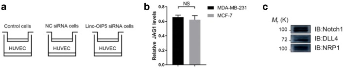
Methods: A trans-well system co-cultured HUVECs with linc-OIP5 knockdown breast cancer cell MDA-MB-231 was utilized to study the proliferation, migration and tube formation abilities of HUVECs and alterations of related signaling indicators in breast cancer cells and their conditioned medium through a series of cell and molecular experiments.

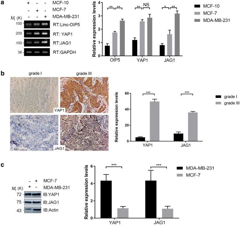
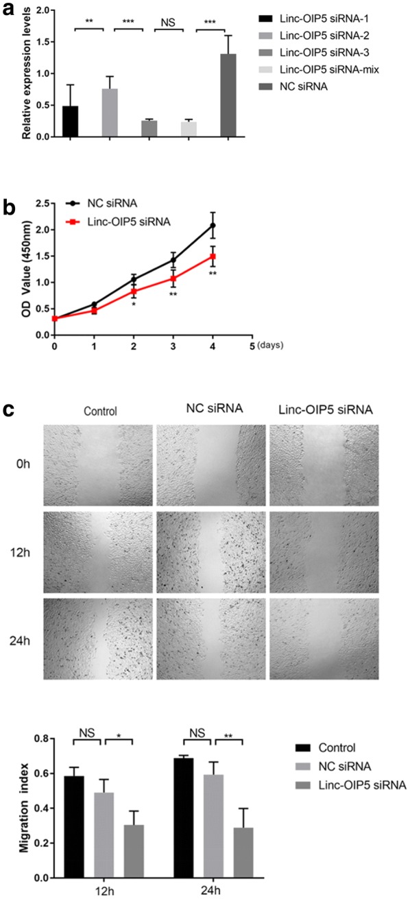
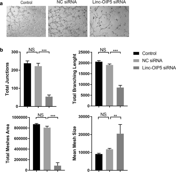
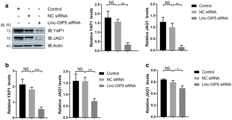
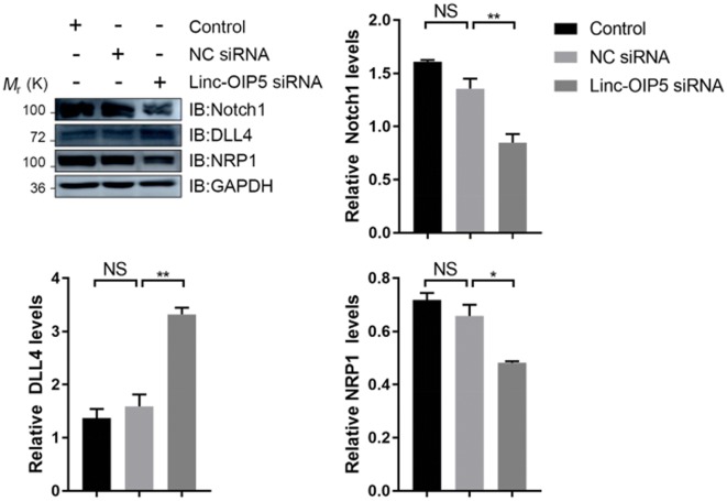
Results: Overexpressed linc-OIP5, YAP1, and JAG1 were found in breast cancer cell lines MCF7 and MDA-MB-231 and the expression levels of YAP1 and JAG1 were proportional to the breast cancer tissue grades. MDA-MB-231 cells with linc-OIP5 knockdown led to weakened proliferation, migration, and tube formation capacity of co-cultured HUVECs. Besides, linc-OIP5 knockdown in co-cultured MDA-MB-231 cells showed downregulated YAP1 and JAG1 expression, combined with a reduced JAG1 level in conditioned medium. Furthermore, a disrupted DLL4/Notch/NRP1 signaling in co-cultured HUVECs were also discovered under this condition.

Conclusion: Hence, linc-OIP5 in MDA-MB-231 breast cancer cells may act on the upstream of the YAP1/Notch/NRP1 signaling circuit to affect proliferation, migration, and tube formation of co-cultured HUVECs in a non-cellular direct contact way through JAG1 in conditioned medium. These findings at least partially provide a new angiogenic signaling circuit in breast cancers and suggest linc-OIP5 could be considered as a therapeutic target in angiogenesis of breast cancers.

Keywords: Angiogenesis; Breast cancer; HUVECs; Linc-OIP5; Signal transduction; Tumor microenvironment.


Conflict of interest statement

The authors declare that they have no competing interests.

Figures

Fig. 1
Fig. 1
Expressions of YAP1 and JAG1 in breast cancer cells and tissues were upregulated. a RT-PCR and its quantification were used to determine the mRNA levels of linc-OIP5, YAP1 and JAG1, which were differentially expressed in normal and cancerous breast cells. b Immunohistochemistry analysis of breast cancer tissue samples confirmed that YAP1 and JAG1 are significantly higher in grade III than grade I and YAP1 in cancer tissues also displayed robust nuclear localization. c Immunoblot analysis and its quantification were performed to check the expression levels of YAP1 and JAG1 in MDA-MB-231 and MCF-7 cells. Fold changes were obtained by normalizing against β-actin. Graphs are means of indicated stained area in 5 samples and error bars indicate s.d. Magnification×200. *P < 0.05, ** P < 0.01, ***P < 0.001
Fig. 2
Fig. 2
MDA-MB-231 cells were adopted to co-culture with HUVECs. a Cell co-culturing simulations: the MDA-MB-231 cells were co-cultured with HUVECs in indirect cell–cell contacts by a trans-well system. b Quantification of JAG1 expression in the conditioned medium of MDA-MB-231 and MCF7 cells. c Immunoblot results of the expression levels of DLL4, Notch1 and NRP1 in HUVECs. NS no statistical difference
Fig. 3
Fig. 3
Knockdown of linc-OIP5 in MDA-MB-231 cells suppressed the proliferation and migration of co-cultured HUVECs in vitro. a Relative expression levels of linc-OIP5 were detected after MDA-MB-231 cells transfected with linc-OIP5 siRNAs. b Knockdown of linc-OIP5 significantly suppressed HUVECs proliferative capacity by CCK-8 assays. c Migration ability of the co-cultured HUVECs after linc-OIP5 knockdown reduced appreciably through wound healing assays. Fold changes were obtained by normalizing against control group. Magnification×40. NS no statistical difference, *P < 0.05, ** P < 0.01
Fig. 4
Fig. 4
Linc-OIP5 knockdown in MDA-MB-231 cells affected angiogenesis capacity of HUVECs in vitro. a HUVECs capillary-like tube formation assay indicated that linc-OIP5 downregulation dramatically reduced the angiogenesis of co-cultured HUVECs. Magnification×40. b Tube formation assay analysis was quantified by the ImageJ software, plotted as the number of total junctions, the lenght of total branching, the area of total meshes, and the size of mean mesh. Fold changes were obtained by normalizing against control group. NS no statistical difference, ** P < 0.01, ***P < 0.001
Fig. 5
Fig. 5
Downregulation of YAP1 and JAG1 was observed in MDA-MB-231 cells with linc-OIP5 knockdown. a Immunoblot confirmed linc-OIP5 siRNA remarkably decreased the protein expressions of YAP1 and JAG1. b qRT-PCR was used to check the mRNA levels of YAP1 and JAG1 after linc-OIP5 knockdown. c Linc-OIP5 siRNA decreased the JAG1 levels in the conditioned medium of MDA-MB-231 cells by ELISA analysis. NS no statistical difference, *P < 0.05, ** P < 0.01, *** P < 0.001
Fig. 6
Fig. 6
Linc-OIP5 knockdown in MDA-MB-231 cells disrupted the Notch1, DLL4 and NRP1 expressions in co-cultured HUVECs. The Notch1, DLL4 and NRP1 expressions were detected by immunoblot using the indicated antibodies. Fold changes were obtained by normalizing against control group. NS no statistical difference, *P < 0.05, ** P < 0.01

Similar articles

Cited by

References

    1. Oon CE, Bridges E, Sheldon H, Sainson RCA, Jubb A, et al. Role of Delta-like 4 in Jagged1-induced tumour angiogenesis and tumour growth. Oncotarget. 2017;8:40115–40131. doi: 10.18632/oncotarget.16969. - DOI - PMC - PubMed
    1. Benedito R, Roca C, Sörensen I, Adams S, Gossler A, et al. The notch ligands Dll4 and Jagged1 have opposing effects on angiogenesis. Cell. 2009;137:1124–1135. doi: 10.1016/j.cell.2009.03.025. - DOI - PubMed
    1. Aspalter IM, Gordon E, Dubrac A, Ragab A, Narloch J, et al. Alk1 and Alk5 inhibition by Nrp1 controls vascular sprouting downstream of Notch. Nat Commun. 2015;6:7264. doi: 10.1038/ncomms8264. - DOI - PMC - PubMed
    1. Guo S, Cheng Y, Ma Y, Yang X. Endothelial progenitor cells derived from CD34+ cells form cooperative vascular networks. Cell Physiol Biochem. 2010;26:679–688. doi: 10.1159/000322335. - DOI - PubMed
    1. Wu N, Nguyen Q, Wan Y, Zhou T, Venter J, et al. The Hippo signaling functions through the Notch signaling to regulate intrahepatic bile duct development in mammals. Lab Invest. 2017;97:843–853. doi: 10.1038/labinvest.2017.29. - DOI - PMC - PubMed

MeSH terms