. 2020:633:203-230.
doi: 10.1016/bs.mie.2019.02.039. Epub 2019 Mar 14.

REX technologies for profiling and decoding the electrophile signaling axes mediated by Rosetta Stone proteins

Affiliations

REX technologies for profiling and decoding the electrophile signaling axes mediated by Rosetta Stone proteins

Marcus J C Long et al. Methods Enzymol. 2020.

Abstract

It is now clear that some cysteines on some proteins are highly tuned to react with electrophiles. Based on numerous studies, it is also established that electrophile sensing underpins rewiring of several critical signaling processes. These electrophile-sensing proteins, or privileged first responders (PFRs), are likely critically relevant for drug design. However, identifying PFRs remains a challenging and unsolved problem, despite the development of several high-throughput methods to ID proteins that react with electrophiles. More importantly, we remain unable to rank how different PFRs identified under different conditions relate to one another, in terms of sensing or signaling capacity. Here we evaluate different methods to assay sensing functions of proteins and discuss these methods in the context of developing a "ranking scheme." Based on theoretical and experimental evidence, we propose that T-REX-the only targeted-electrophile delivery tool presently available-is a reliable method to rank PFRs. Finally, we address to what extent electrophile sensing and downstream signaling are correlated. Based on our current data, we observe that such behaviors are indeed correlated. It is our hope that through this manuscript researchers from various arms of the stress signaling fields will focus on developing a quantitative understanding of precision electrophile labeling.

Keywords: Electrophile signaling; G-REX; Privileged first responders; Ranking reactivity; Redox-regulated pathways; Stress response; T-REX.


Figures

Fig. 1.
Fig. 1.
Representative methods of high-throughput (HT) identification (ID) of RES-sensing proteins. (A) Competitive activity-based protein profiling (ABPP) adapted to target ID of HNE-sensing proteins. Treatment of lysates [originating from cells treated with either HNE (100 μM, 1 h) or DMSO] with alkynylated activity probe (alkynylated iodoacetamide; 100 μM, 1 h), followed by Click coupling with light or heavy TEV-cleavable biotin tags (Plight and Pheavy), respectively, streptavidin enrichment, and downstream MS/MS analysis, altogether enable indirect report of HNE sensitivity (Wang, Weerapana, Blewett, & Cravatt, 2014). (B) ABPP ranks proteins by “R value,” a ratio of heavy/light probe modification (POI-Plight/POI-Pheavy) that serves as a readout of the intrinsic kinetics of HNE labeling by a specific protein (kPCI), assuming constant POI-concentration, [POI] (Wang et al., 2014). (C) ID of HNE sensors built on utilization of pseudo-endogenous generation of HNE from alkynylated precursors. Incorporation of linoleic acid (LA) into phospholipids, followed by Kdo2-lipidA-activated autoxidation (see text for details), reveals potential key players in stress-response (Beavers et al., 2017).
Fig. 2.
Fig. 2.
G-REX mines native RES-sensors with high spatiotemporal resolution and controlled RES chemotype and dosage. G-REX is built on direct capture of native sensors in vivo. Following treatment of live specimens with the specific photocaged REX-probe and washing away the unbound excess probe, the concentration of RES released in the cell (or a specific organelle/tissue), upon subsequent photouncaging at a preordained time, is at maximum equal to the concentration of HaloTag in vivo (Liu, Long, & Aye, 2019; Zhao et al., 2018). Locale of the RES release is determined by where Halo is expressed (also see Fig. 3). G-REX thus more closely mimics native electrophilic signaling and offers a user-controlled RES-sensor ID. By utilizing a specific RES/covalent-ligand such as HNE as a chemical-signal input, a quantitative G-REX output can be obtained ([POI-HNE], where POI is the putative protein sensor). Further downstream work is required to more accurately assess the relative rates of RES-reactivity across the top hits, since the output is affected by both intrinsic kinetic properties kn and endogenous concentration of the individual sensor protein-of-interest [POI]n. as well as [HNE] at a given time t.
Fig. 3.
Fig. 3.
Expanding G-REX toward locale-specific sensing and target ID in complex live models. (A) REX-probes have been shown to be compatible with complex living systems such as Danio rerio (Long et al., 2017) and C. elegans (Long et al., 2018), rendering G-REX platform amenable to mining novel sensors in these animals. One of the on-going applications of G-REX involves locale-specific mining of RES-sensors at the live organismal level; e.g., organelle-specific HaloTag expression [mitochondrial outer membrane (OM) localization via OM-anchoring domain of Tom70, OM-translocase from Saccharomyces cerevisiae] can be achieved in complex model organisms such as C. elegans. Scale bars are 50 μm. (B) G-REX integrating localized expression of Halo (such as to mitochondria, right) explores region-specific RES-sensors that may have otherwise been missed through ubiquitous/whole-cell HaloTag expression (left). Concentric shaded rings in the diagram depict different diffusion distances for a RES such as HNE. Note: for locale-specific G-REX, these distances should be as low as possible: e.g., for mitochondria (at cross-section diameters reportedly up to 1 μm), longer linear diffusion distances would lead to drastic increases in the treated cell volume, reducing organelle specificity. Panel (A): Reprinted (adapted) with permission from Long, M. J. C., Urul, D. A., Chawla, S., Lin, H. Y., Zhao, Y., Haegele, J. A., et al. (2018). Precision electrophile tagging in Caenorhabditis elegans. Biochemistry, 57(2), 216–220. doi:https://doi.org/10.1021/acs.biochem.7b00642 (web archive link). Copyright (2017) American Chemical Society.
Fig. 4.
Fig. 4.
T-REX is a unique and necessary tool for directly interrogating and validating signaling outputs exclusively arising from POI-specific RES-modifications. (Left) Bulk HNE exposure to cells/animals—while representing a rapid and facile means to ID candidate RES-sensors—render challenges in studying downstream consequences of specific sensor RES-modification. These difficulties are in part due to: off-target RES-binding and associated toxicity and secondary responses, etc., following RES-saturation of first-responding sensor cysteines; uncontrolled conversion of RES in cells into other reactive metabolites that can adduct additional proteins; and perturbation of cellular redox balance. (Right) T-REX enables selective labeling of the target protein-of-interest (POI) in the cell or animals, through controlled release of RES from a photocaged precursor bound to Halo fused to POI. T-REX enables a clearer insight into a given POI—RES-pair-specific modulation of signaling output as well as the kinetics of RES-sensor modification. Note: The REX-probe used in G-REX (see Fig. 2) and T-REX is identical for a given RES of interest.
Fig. 5.
Fig. 5.
T-REX analyses HNE-sensing kinetics of individual POIs (Left). In the T-REX model, the release of HNE and subsequent covalent modification happens in a pseudo-intramolecular fashion within the solvent cage surrounding Halo-POI. As such, the observed rate of this reaction (rnterm or rcterm; “nterm” and “cterm” designating, respectively, N- or C-terminal Halo-fusion) are independent of POI (here, Keap1) or released-RES concentration, and are instead governed by the off-rate of the released RES, here HNE (diffusing into the environment; knoff) and the on-rate of covalent modification of the POI (knon) (Parvez et al., 2016). (Right) In a bimolecular system (so-called split system, where Halo and POI are separately expressed), this is not the case (Lin, Haegele, Disare, Lin, & Aye, 2015). Here, both the sensor-POI (Keap1) and the released RES can vary in concentration, and the rate itself (rbm; “bm” designating bimolecular) is concentration-dependent. Further, release of the RES into the cell from Halo enables the measured rate to be influenced by HNE-binding to off-targets. The “Cy5 gel” illustrates a representative in-gel fluorescence data set from T-REX targeting efficiency analysis, whereby labeling is evaluated by Cy5-azide click coupling that reports on alkyne signal (either within the photocaged probe or the released RES that, respectively, bind Halo and RES-sensor-protein). The blots below the Cy5 gel correspond to western blots confirming the presence of indicated protein in the lysate. Lane 1 corresponds to data originating from cells expressing the Halo-Keap1 that were subjected to full T-REX conditions, indicating HNE(alkyne)-modification of Keap1 (note: “post TEV cleavage” designates samples that were subjected to TEV-protease-assisted separation of Halo and Keap1 post cell lysis prior to Click coupling with Cy5-azide). Lanes 2 and 3 correspond to a bimolecular system (i.e., the “split” system) where Halo and Keap1 are expressed separately (each fused to GFP) in cells. Data from these samples show no HNE(alkyne)-modification of Keap1. Lane 4 corresponds to the same bimolecular system harvested without any photouncaging, demonstrating the “photocaged-probe-loaded” HaloTag. (Bottom) The measured output of T-REX kinetics is largely intrinsic to the POI itself (specifically kon), enabling the “targeting efficiency” parameter derived from T-REX analysis to be used for “ranking” of differential sensitivity to a given RES (if k1on < k2on, then corresponding r1 < r2; thus, rates can be used to rank sensors) (see text for details). Reprinted (adapted) with permission from Lin, H. Y., Haegele, J. A., Disare, M. T., Lin, Q., & Aye, Y. (2015). A generalizable platform for interrogating target- and signal-specific consequences of electrophilic modifications in redox-dependent cell signaling. Journal of the American Chemical Society, 137(19), 6232–6244. doi:10.1021/ja5132648. Copyright (2015) American Chemical Society.
Fig. 6.
Fig. 6.
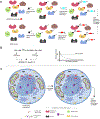
A case study of the recent discovery pipeline enabled by REX technologies. (A) Ube2V2 was uncovered as a sensing candidate using inaugural G-REX approach (Zhao et al., 2018). Through T-REX-assisted evaluation of relative RES-sensitivity (i.e., targeting efficiency/site-occupancy, see text for discussion), Ube2v2 was less sensitive to HNE than Keap1. (B) Ube2V2 translates RES modification of a sensing cysteine (black; distinguished from the red catalytic cysteine on Ube2N) into a ubiquitin code through allosteric stimulation of Ube2N activity, consequently modulating DNA damage response signaling. This type of signal translation and propagation enables Ube2V2 to act as a “Rosetta Stone” proteins, converting the chemical language of electrophiles to that of canonical signal, ubiquitin (Zhao et al., 2018). This mode-of-action by HNE on a specific protein functions differently to the direct modulation of Keap1 function.
Fig. 7.
Fig. 7.
T-REX ranks relative RES-sensitivity across RES-sensor POIs and G-REX unearths “Rosetta Stone” sensing and RES-signal translation. Differential RES sensitivity as a possible insight into the mechanisms of downstream signal propagation. (Top) Higher-ranking HNE sensors (i.e., those judged to have higher relative targeting efficiency/target occupancy derived by T-REX analysis such as Keap1) (Lin et al., 2015; Long & Aye, 2016; Parvez et al., 2015) and Akt3 (Long et al., 2017; Parvez et al., 2016), directly influence downstream responses as “first-responder” sensors. (Bottom) By contrast, lower-ranking sensors such as Ube2V2 mediate sensing through activation of a binding-partner (Ube2N in this case) in an allosteric fashion (Zhao et al., 2018).

Similar articles

Cited by

References

    1. Amalraj A, Pius A, Gopi S, Gopi S, 2017. Biological activities of curcuminoids, other biomolecules from turmeric and their derivatives—A review. Journal of Traditional and Complementary Medicine 7 (2), 205–233. 10.1016/j.jtcme.2016.05.005. - DOI - PMC - PubMed
    1. Anand DAV, Arulmoli R, Parasuraman S, 2016. Overviews of biological importance of quercetin: A bioactive flavonoid. Pharmacognosy Reviews 10 (20), 84–89. 10.4103/0973-7847.194044. - DOI - PMC - PubMed
    1. Beavers WN, Rose KL, Galligan JJ, Mitchener MM, Rouzer CA, Tallman KA, et al., 2017. Protein modification by endogenously generated lipid electrophiles: Mitochondria as the source and target. ACS Chemical Biology 12 (8), 2062–2069. 10.1021/acschembio.7b00480. - DOI - PMC - PubMed
    1. Blatnik M, Frizzell N, Thorpe SR, Baynes JW, 2008. Inactivation of glyceraldehyde-3-phosphate dehydrogenase by fumarate in diabetes: Formation of S-(2-succinyl)cysteine, a novel chemical modification of protein and possible biomarker of mitochondrial stress. Diabetes 57 (1), 41–49. 10.2337/db07-0838. - DOI - PMC - PubMed
    1. Blewett MM, Xie J, Zaro BW, Backus KM, Altman A, Teijaro JR, et al., 2016. Chemical proteomic map of dimethyl fumarate-sensitive cysteines in primary human T cells. Science Signaling 9 (445), rs10 10.1126/scisignal.aaf7694. - DOI - PMC - PubMed

Publication types

LinkOut - more resources