. 2020 Feb 25;117(8):4300-4309.
doi: 10.1073/pnas.1920021117. Epub 2020 Feb 11.

Nucleolar localization of RAG1 modulates V(D)J recombination activity

Affiliations

Nucleolar localization of RAG1 modulates V(D)J recombination activity

Ryan M Brecht et al. Proc Natl Acad Sci U S A. .

Abstract

V(D)J recombination assembles and diversifies Ig and T cell receptor genes in developing B and T lymphocytes. The reaction is initiated by the RAG1-RAG2 protein complex which binds and cleaves at discrete gene segments in the antigen receptor loci. To identify mechanisms that regulate V(D)J recombination, we used proximity-dependent biotin identification to analyze the interactomes of full-length and truncated forms of RAG1 in pre-B cells. This revealed an association of RAG1 with numerous nucleolar proteins in a manner dependent on amino acids 216 to 383 and allowed identification of a motif required for nucleolar localization. Experiments in transformed pre-B cell lines and cultured primary pre-B cells reveal a strong correlation between disruption of nucleoli, reduced association of RAG1 with a nucleolar marker, and increased V(D)J recombination activity. Mutation of the RAG1 nucleolar localization motif boosts recombination while removal of the first 215 amino acids of RAG1, required for efficient egress from nucleoli, reduces recombination activity. Our findings indicate that nucleolar sequestration of RAG1 is a negative regulatory mechanism in V(D)J recombination and identify regions of the RAG1 N-terminal region that control nucleolar association and egress.

Keywords: B cell development; RAG1; V(D)J recombination; nucleolus; proximity-dependent biotin identification.


Conflict of interest statement

The authors declare no competing interest.

Figures
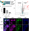
Fig. 1.
Fig. 1.
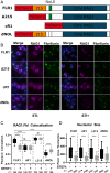
BioID reveals nucleolar association of RAG1. (A) Schematic of the three BioID constructs N-terminally fused to RAG1 and the control construct fused to a NLS. (B) Volcano plot comparing enriched proteins between core RAG1 and Δ215. P value is on a −Log10 scale. Nucleolar proteins are shown as red squares. (C) Bar graph showing the number of nucleolar proteins found enriched with each RAG1 construct compared to the control. (D) Confocal images of HEK293T cells transiently transfected with mCherry fused to either Δ215 or core RAG1, imaged 24 h after transfection, showing colocalization of Δ215 with the nucleolar marker GFP-fibrillarin. Representative of three independent experiments.
Fig. 2.
Fig. 2.
Nucleolar localization of RAG1 is dynamic and dependent on the recombination state of the cell. (A) Schematic of RAG1 fusion proteins used for analysis. (B) Confocal images from vAbl cells constitutively expressing GFP-fused fibrillarin and doxycycline-inducible mCherry-fused RAG1. Cells were treated with doxycycline to induce RAG1 expression for 16 h and, when stated, treated with 5 µM STI-571 for 4 h prior to being mounted and fixed. Representative of three independent experiments. (C) Colocalization analysis between RAG1 and fibrillarin (Fbl) before and after STI-571 treatment. Pearson correlation was calculated from individual cells and plotted with whiskers at 10th to 90th percentile. Statistical significance was determined by ANOVA. ****P < 0.0001; NS, not significant. (D) Quantitation of nucleolar size by pixel area before and after STI-571 treatment. Individual nucleoli plotted with whiskers at 10th to 90th percentile. Statistical significance was determined by ANOVA. ****P < 0.0001.
Fig. 3.
Fig. 3.
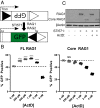
Recombination efficiency is regulated by nucleolar localization of RAG1. (A) Schematic of pMGInv inversional GFP reporter. Black and white triangles represent 23RSS and 12RSS, respectively. (B) Recombination efficiency assay using pMGInv reporter in WT and core RAG1 vAbl cells. Cells were induced for 48 h with 5 µM STI-571 with varying amounts of actD and analyzed via flow cytometry for GFP+ cells. Statistical significance was determined by ANOVA. ****P < 0.0001; NS, not significant. (C) Western blot of RAG1 from vAbl cells treated with 5 µM STI-571 and/or 2 nM actD for 48 h. ActD does not influence RAG1 expression levels. Representative of two independent experiments.
Fig. 4.
Fig. 4.
Nucleolar morphology is affected by actD treatment. (A) Confocal images from vAbl cells constitutively expressing GFP-fused fibrillarin and doxycycline-inducible mCherry-fused RAG1. Cells were treated with doxycycline to induce RAG1 expression for 16 h and with 2 nM actD for 4 h prior to being mounted and fixed. Representative of three independent experiments. (B) Colocalization analysis between RAG1 and fibrillarin (Fbl) before and after actD treatment. (C) Quantitation of nucleolar size by pixel area before and after actD treatment. Individual nucleoli plotted with whiskers at 10th to 90th percentile. Statistical significance was determined by ANOVA. ****P < 0.0001.
Fig. 5.
Fig. 5.
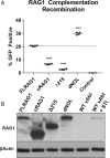
Functional comparison of RAG1 truncations reveals distinct roles for different parts of the RAG1 NTD. (A) Recombination efficiency from a clonal RAG1−/− RAG2+/+ vAbl cell line transduced with different forms of RAG1. At 72 h postinfection, cells were analyzed via flow cytometry for GFP+ cells. Statistical significance was determined by ANOVA. ****P < 0.0001. (B) Western blot showing expression levels of RAG1 from infected RAG1-complementation lines. WT vAbl cells uninduced and induced with STI-571 shown as control. Representative of three independent experiments.
Fig. 6.
Fig. 6.
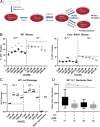
Recombination efficiency in primary pre-B cells is altered by actinomycin D. (A) Schematic showing culture conditions during assay. (B) pMGInv recombination assay of homogenized pre-B cells from WT and core RAG1 mice. Cells were analyzed for GFP+ cells after 48 h with/without IL7 in various concentration of actD. Representative of two independent experiments. Statistical significance was determined by ANOVA. **P < 0.01; ***P < 0.001; ****P < 0.0001; NS, not significant. (C) ddPCR assay for intact Jκ2, as assessed by the decrease in PCR amplification resulting from Igκ cleavage and recombination. The signal for intact Jκ2 alleles was measured, normalized to an amplicon from the gene RPP30, and then subtracted from 2 to yield the apparent level of Jκ2 cleavage. Genomic DNAs from kidney and B220+ splenocytes were used as no cleavage and high cleavage controls, respectively. For IL7 culture data, each data point represents the mean of either two or eight technical replicates performed for two independent samples. For spleen and kidney, each data point represents the mean of four technical replicates for two independent samples. Statistical significance was determined by ANOVA. **P < 0.01; ****P < 0.0001. (D) Quantitation of nucleolar size by pixel area in pre-B cells untreated and treated with IL7 and/or 2 nM actD for 48 h. Individual nucleoli plotted with whiskers at 10th to 90th percentile. Statistical significance was determined by ANOVA. ****P < 0.0001.

References

    1. Tonegawa S., Somatic generation of antibody diversity. Nature 302, 575–581 (1983). - PubMed
    1. Schatz D. G., Oettinger M. A., Baltimore D., The V(D)J recombination activating gene, RAG-1. Cell 59, 1035–1048 (1989). - PubMed
    1. Oettinger M. A., Schatz D. G., Gorka C., Baltimore D., RAG-1 and RAG-2, adjacent genes that synergistically activate V(D)J recombination. Science 248, 1517–1523 (1990). - PubMed
    1. Schatz D. G., Ji Y., Recombination centres and the orchestration of V(D)J recombination. Nat. Rev. Immunol. 11, 251–263 (2011). - PubMed
    1. Gellert M., V(D)J recombination: RAG proteins, repair factors, and regulation. Annu. Rev. Biochem. 71, 101–132 (2002). - PubMed

Publication types