. 2020 Feb 25;117(8):4281-4291.
doi: 10.1073/pnas.1909814117. Epub 2020 Feb 11.

Decision between mitophagy and apoptosis by Parkin via VDAC1 ubiquitination

Affiliations

Decision between mitophagy and apoptosis by Parkin via VDAC1 ubiquitination

Su Jin Ham et al. Proc Natl Acad Sci U S A. .

Abstract

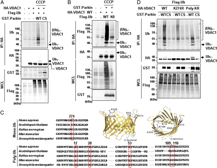
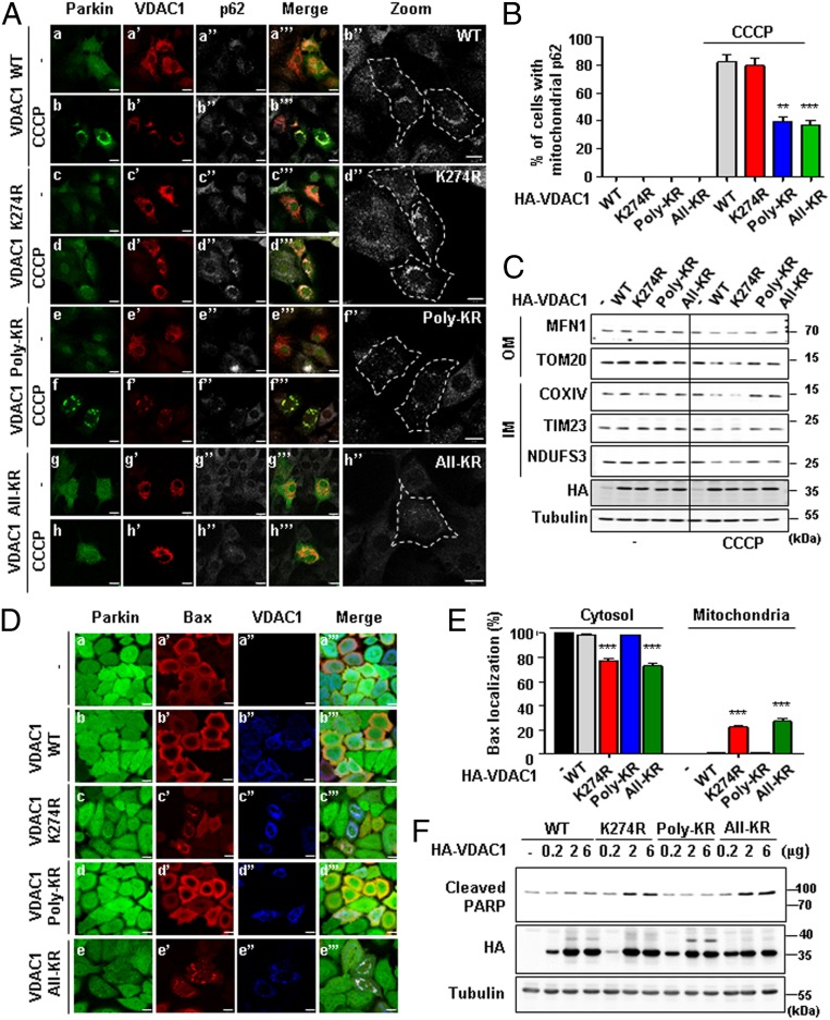
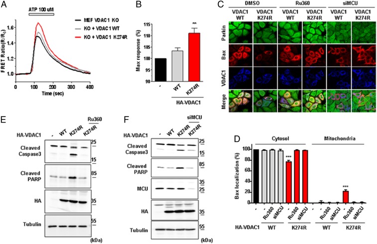
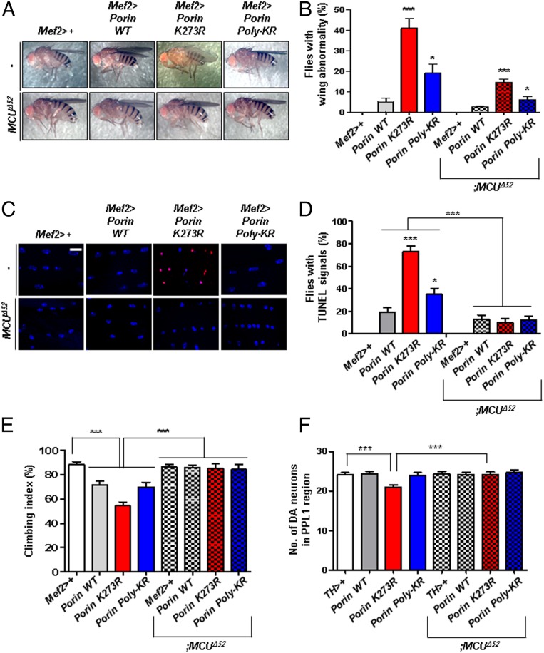
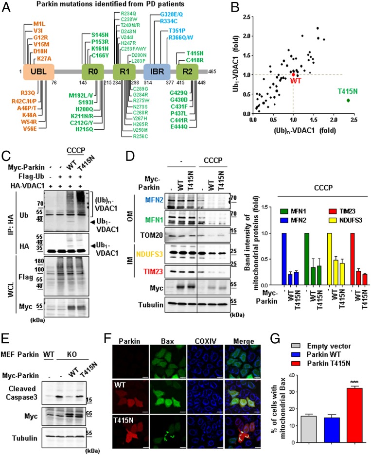
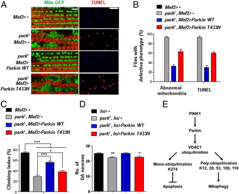
VDAC1 is a critical substrate of Parkin responsible for the regulation of mitophagy and apoptosis. Here, we demonstrate that VDAC1 can be either mono- or polyubiquitinated by Parkin in a PINK1-dependent manner. VDAC1 deficient with polyubiquitination (VDAC1 Poly-KR) hampers mitophagy, but VDAC1 deficient with monoubiquitination (VDAC1 K274R) promotes apoptosis by augmenting the mitochondrial calcium uptake through the mitochondrial calcium uniporter (MCU) channel. The transgenic flies expressing Drosophila Porin K273R, corresponding to human VDAC1 K274R, show Parkinson disease (PD)-related phenotypes including locomotive dysfunction and degenerated dopaminergic neurons, which are relieved by suppressing MCU and mitochondrial calcium uptake. To further confirm the relevance of our findings in PD, we identify a missense mutation of Parkin discovered in PD patients, T415N, which lacks the ability to induce VDAC1 monoubiquitination but still maintains polyubiquitination. Interestingly, Drosophila Parkin T433N, corresponding to human Parkin T415N, fails to rescue the PD-related phenotypes of Parkin-null flies. Taken together, our results suggest that VDAC1 monoubiquitination plays important roles in the pathologies of PD by controlling apoptosis.

Keywords: PINK1; Parkin; Parkinson disease; VDAC1; apoptosis.


Conflict of interest statement

The authors declare no competing interest.

Figures

Fig. 1.
Fig. 1.
Mono- and polyubiquitination of VDAC1 are dependent on Parkin and PINK1 activity. (A, B, and D) Ubiquitination assays for VDAC1 by Parkin. HEK293T cells were transfected with indicated constructs and treated with or without 20 µM CCCP for 4 h. Whole-cell lysates (WCL) were immunoprecipitated (IP) and analyzed for IB analyses with indicated antibodies. The monoubiquitination band was marked on the right side and the polyubiquitination bands were marked by a bracket. Parkin WT and CS (C431S) mutants were expressed and compared in A and D. Flag-tagged ubiquitin WT and K0 (K6, 11, 27, 29, 33, 48, and 63R mutant) were expressed in B. VDAC1 WT and two ubiquitination mutants (VDAC1 K274R and Poly-KR) were expressed and also examined for Parkin-dependent ubiquitination assays in D. (C) Aligned VDAC1 protein sequences of human (VDAC1, P21796.2), Arabidopsis (VDAC1, NP_186777.1), rat (VDAC1, NP_112643.1), mouse (VDAC1 isoform 2, NP_035824.1), and Drosophila (Porin isoform E, NP_001260365.1) with indication of the conserved lysine residues in red boxes. Three-dimensional human VDAC1 protein structures, Protein Data Bank ID code 5JDP (P21796), were additionally used to predict the location of mono- and polyubiquitination sites.
Fig. 2.
Fig. 2.
Poly- and monoubiquitination on VDAC1 determine the fate of mitochondria to mitophagy or apoptosis, respectively. (A) Confocal images of the translocation of p62/SQSTM1 to mitochondria. VDAC1 KO MEF cells were transfected with HA-tagged VDAC1 WT, K274R, Poly-KR, or All-KR and treated with 30 µM CCCP for 8 h. Subcellular localizations of YFP-Parkin (green), VDAC1 (red), and p62/SQSTM1 (gray) were observed. (Scale bars, 20 µm.) (B) Percentage of the cells with p62/SQSTM1 localized to the mitochondria in A (n > 500 cells from three independent experiments). Data were analyzed by ANOVA Tukey test. **P < 0.01; ***P < 0.001. (C) Concentrations of mitochondrial proteins from the outer membrane (OM) and inner membrane (IM) in VDAC1 KO MEF cells with indicated VDAC1 proteins transfected for 48 h and treated with or without 30 µM CCCP for 16 h. (D) Confocal images of Bax translocated to the mitochondria. Subcellular localizations of GFP-Parkin (green), Bax (red), and VDAC1 proteins (blue) were observed. (Scale bars, 20 µm.) (E) Percentage of the cells with Bax localized to mitochondria in D (n > 500 cells from three independent experiments). Data were analyzed by one-way ANOVA with Tukey multiple-comparison test and are presented as means ± SD. ***P < 0.001. (F) HeLa cells stably expressing GFP-Parkin were transfected with indicated VDAC1 proteins in a dose-dependent manner and analyzed by immunoblotting to examine indicated proteins.
Fig. 3.
Fig. 3.
Monoubiquitination-deficient VDAC1 induces apoptosis by increasing mitochondrial calcium uptake. (A and B) Mitochondrial calcium dynamics in VDAC1 KO MEF cells. VDAC1 KO MEF cells were transfected with HA-tagged VDAC1 WT or K274R and cotransfected with 4mitD3, a mitochondrial calcium indicator, for 48 h. Cells were then treated with 100 µM ATP for 3 min and were monitored for the changes in 4mitD3 fluorescence. (B) Maximum mitochondrial calcium uptake for VDAC1 KO MEF cells expressing VDAC1 WT or K274R in A. All of the values were normalized to the basal level of VDAC1 KO MEF cells transfected with empty vector (−) (n = 50 to ∼70 cells). Data were analyzed by ANOVA Tukey test. **P < 0.05. (C and D) Confocal images of Bax translocated to the mitochondria. (C) Flag-tagged Bax (red) and HA-tagged VDAC1 WT or K274R (blue) were transfected and immune-stained with Flag and HA antibodies in HeLa cells stably expressing GFP-Parkin (green). Cells were treated with or without 0.5 mM Ru360 for 8 h or transfected with MCU siRNA. (Scale bars, 20 µm.) (D) Percentage of cells with Bax localized in the cytosol or the mitochondria shown in C (n > 500 cells from three independent experiments). Data were analyzed by one-way ANOVA with Tukey multiple-comparison test and are presented as means ± SD. ***P < 0.001. (E and F) HA-tagged VDAC1 WT or K274R was transfected in HeLa cells stably expressing GFP-Parkin. Cells were treated with or without 0.5 mM Ru360 for 8 h in E or transfected with MCU siRNA in F. Cell lysates were analyzed by immunoblotting to examine indicated proteins.
Fig. 4.
Fig. 4.
All phenotypes related to PD in Porin K273R flies are rescued by suppressing MCU. (A and B) Defects in the wing posture of Porin K273R flies. Transgenic flies expressing Porin WT, K273R, or Poly-KR using Mef2-Gal4 driver were crossed with MCU null flies (MCUΔ52). (B) Percentage of the flies with defective wing postures in A (n = 300 from three independent experiments). (C and D) TUNEL signals for apoptosis (red) and 4′,6-diamidino-2-phenylindole staining for the nucleus (blue) in the thoraces of transgenic flies. (Scale bar, 5 µm.) (D) Percentage of the flies with TUNEL signals in C (n = 100 from 10 independent experiments). (E) Percentage of the transgenic flies with defective climbing ability (n = 100 from 10 independent experiments). (F) Numbers of DA neurons in the brain PPL1 region (n = 10). All quantifications were analyzed by one-way ANOVA with Tukey multiple-comparison test and are presented as means ± SD. ***P < 0.001; *P < 0.05.
Fig. 5.
Fig. 5.
Impaired monoubiquitination of VDAC1 by Parkin T415N leads to apoptosis. (A) Parkin PD patient mutations in the domains of Parkin protein. Listing from the N-terminal to the C-terminal region, Parkin consists of UBL, really-interesting-new-gene (RING) 0, RING1, in-between RING (IBR), and RING2 domains. (B) Summarized results of the ubiquitination assays for VDAC1 by 63 different Parkin PD patient mutants. Mono- and polyubiquitination activities of these Parkin mutants were normalized to the mono- and polyubiquitination of VDAC1 by Parkin WT (red). The Parkin T415N mutant (green) showed significant reduction in the monoubiquitination of VDAC1. We quantitated band intensities of ubiquitination on VDAC1 using Image J program. (C) Ubiquitination assays on the Parkin T415N mutant. Monoubiquitination of VDAC1 is indicated by arrows, and the bracket represents the polyubiquitination. IP, immunoprecipitated; WCL, whole-cell lysates. (D) Mitochondrial proteins analyzed by immunoblotting. HeLa cells were expressed with Myc-tagged Parkin WT or T415N with or without 20 µM CCCP for 12 h. A quantitation graph shows the protein band intensity of MFN2 (blue), MFN1 (green), NDUFS3 (yellow), and TIM23 (red) normalized to tubulin from CCCP-treated cells. Data were obtained from two independent experiments and are shown as means ± SD. (E) Parkin KO MEF cells expressing Myc-tagged Parkin WT or T415N were compared with Parkin WT MEF cells. Whole-cell lysates were analyzed for immunoblotting to detect indicated proteins. (F) Confocal images of Bax translocated to the mitochondria. HeLa cells were transfected with Myc-tagged Parkin WT or T415N and treated with 1 mM H2O2 for 6 h. Subcellular localizations of Bax (green), COXIV (blue), and Parkin (red) were examined by confocal microscopy. (Scale bars, 20 µm.) (G) Percentage of cells with Bax in mitochondria in F (n > 450 cells from three independent experiments). Data were analyzed by ANOVA Tukey test. ***P < 0.001
Fig. 6.
Fig. 6.
Drosophila Parkin T433N cannot rescue the PD-related phenotypes of the park1 mutant. (A) Mitochondrial morphology and TUNEL signals in the thoraces of Parkin-null flies (park1) expressing Parkin WT or T433N using the Mef2-Gal4 driver. Mitochondrial morphology (green) and phalloidin staining (red) to indicate muscle fiber are shown (Left). TUNEL signals (red) and 4′,6-diamidino-2-phenylindole staining (blue) are shown (Right). (Scale bars, 5 μm.) (B) Percentage of flies with defective mitochondrial morphology or TUNEL signals (n = 100 from 10 independent experiments). (C) Percentage of flies with defective climbing ability (n = 100 from 10 independent experiments). (D) Numbers of DA neurons in the PPL1 region (n = 10). All quantifications were analyzed by one-way ANOVA with Tukey multiple-comparison test and are presented as means ± SD. *P < 0.05; **P < 0.01; ***P < 0.001. (E) A schematic model of mitophagy and apoptosis regulated by two types of ubiquitination on VDAC1 in the PINK1–Parkin pathway.

References

    1. Baba Y., et al. , Phenotypic commonalities in familial and sporadic Parkinson disease. Arch. Neurol. 63, 579–583 (2006). - PubMed
    1. Bonifati V., et al. , Mutations in the DJ-1 gene associated with autosomal recessive early-onset parkinsonism. Science 299, 256–259 (2003). - PubMed
    1. Hague S., et al. , Early-onset Parkinson’s disease caused by a compound heterozygous DJ-1 mutation. Ann. Neurol. 54, 271–274 (2003). - PubMed
    1. Hatano Y., et al. , Novel PINK1 mutations in early-onset parkinsonism. Ann. Neurol. 56, 424–427 (2004). - PubMed
    1. Kitada T., et al. , Mutations in the parkin gene cause autosomal recessive juvenile parkinsonism. Nature 392, 605–608 (1998). - PubMed

Publication types

MeSH terms