

Central compact osteoma of mandibular condyle
- PMID: 32047087
- PMCID: PMC7035841
- DOI: 10.1136/bcr-2019-233082
Central compact osteoma of mandibular condyle
Abstract
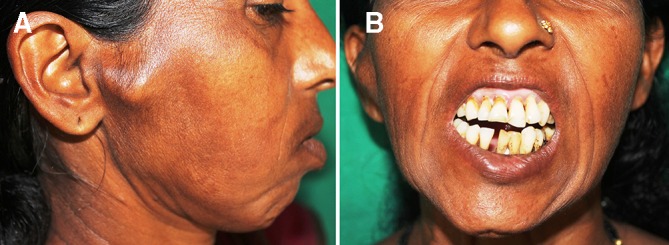
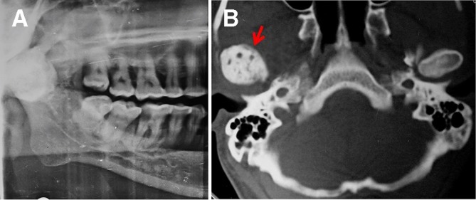
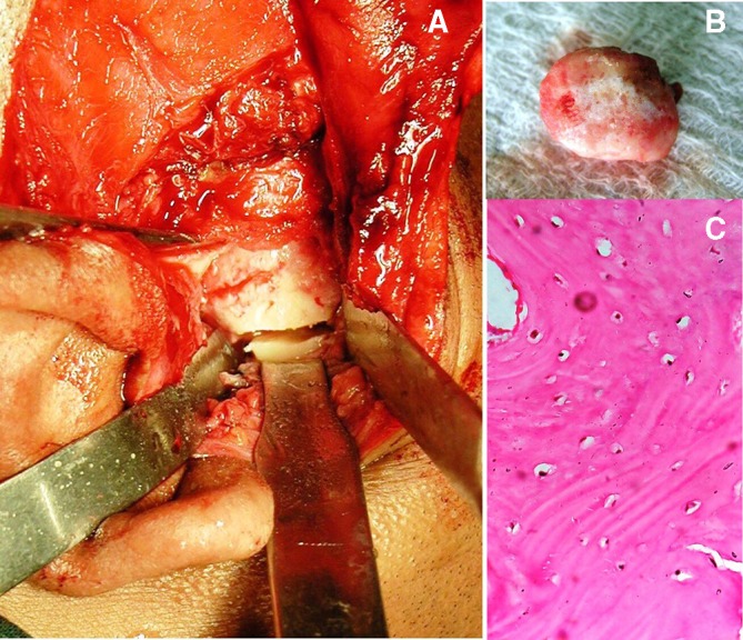
Osteomas are bone tumours arising from the cortical or medullary bones of craniofacial skeleton. Involvement of frontal bone and paranasal sinuses is more frequent than jaw bones. Jaw osteomas are slow growing benign lesions, which are usually asymptomatic or present as painless swelling. Those involving mandibular condyle are relatively rare and result in significant functional and aesthetic disturbances. This paper reports a case of solitary central compact osteoma of mandibular condyle in an adult Indian female patient. A comprehensive review of previously published reports is also presented.
Keywords: dentistry and oral medicine; oral and maxillofacial surgery; surgery.
© BMJ Publishing Group Limited 2020. No commercial re-use. See rights and permissions. Published by BMJ.
Conflict of interest statement
Competing interests: None declared.
Figures
Similar articles
-
Giant mandibular condyle osteoma.J Craniofac Surg. 2011 May;22(3):1147-9. doi: 10.1097/SCS.0b013e318210baee. J Craniofac Surg. 2011. PMID: 21586974
-
Osteoma of the mandibular condyle: report of a case with 5-year follow-up.Mil Med. 2005 Feb;170(2):117-20. Mil Med. 2005. PMID: 15782830
-
Endoscopic resection of a mandibular body and condylar osteoma.Minim Invasive Ther Allied Technol. 2008;17(5):323-5. doi: 10.1080/13645700802389964. Minim Invasive Ther Allied Technol. 2008. PMID: 18855202
-
Osteoma of the mandibular condyle: a rare case report and review of the literature.J Stomatol Oral Maxillofac Surg. 2019 Dec;120(6):584-587. doi: 10.1016/j.jormas.2019.01.013. Epub 2019 Jan 25. J Stomatol Oral Maxillofac Surg. 2019. PMID: 30685345 Review.
-
Rare benign tumors of the mandibular condyle: report of 2 cases and literature review.J Oral Maxillofac Surg. 2007 Sep;65(9):1830-5. doi: 10.1016/j.joms.2006.06.262. J Oral Maxillofac Surg. 2007. PMID: 17719407 Review. No abstract available.
Cited by
-
Central ossifying fibroma of mandible.BMJ Case Rep. 2020 Dec 28;13(12):e239286. doi: 10.1136/bcr-2020-239286. BMJ Case Rep. 2020. PMID: 33372024 Free PMC article.
-
Craniofacial Osteomas: From Diagnosis to Therapy.J Clin Med. 2021 Nov 27;10(23):5584. doi: 10.3390/jcm10235584. J Clin Med. 2021. PMID: 34884284 Free PMC article. Review.
References
Publication types
MeSH terms
LinkOut - more resources
Full Text Sources
Medical