. 2020 Feb 11;10(1):2357.
doi: 10.1038/s41598-020-59157-2.

Effects of corneal crosslinking on corneal shape stabilization after orthokeratology

Affiliations

Effects of corneal crosslinking on corneal shape stabilization after orthokeratology

Chimei Liao et al. Sci Rep. .

Abstract

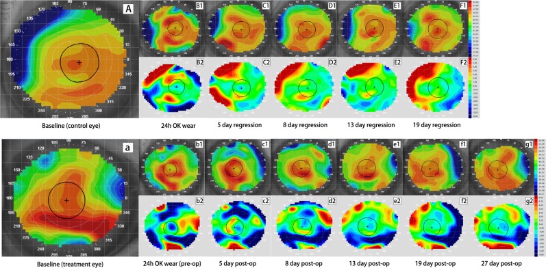
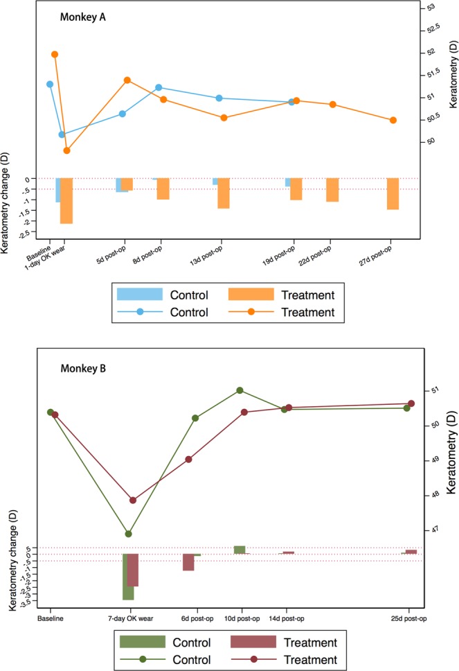
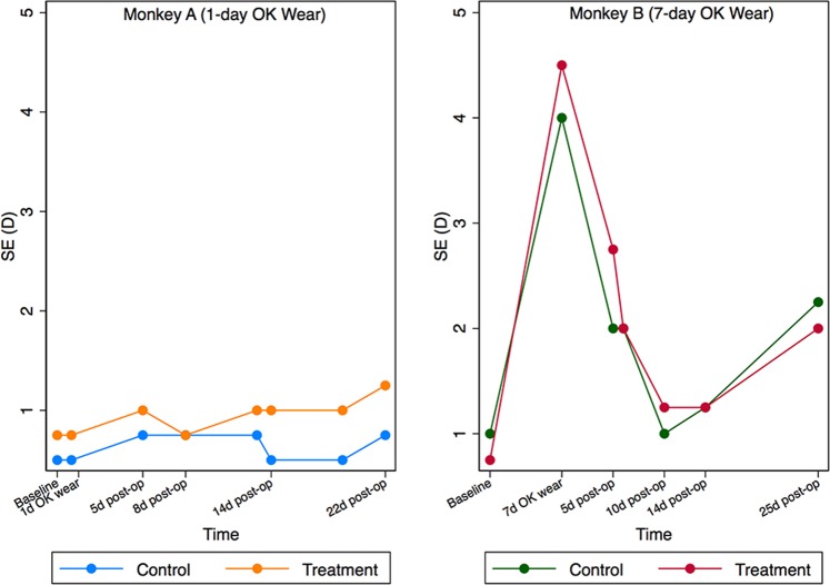
Orthokeratology (Ortho-K) works to reshape cornea and is the only non-surgical way to enable vision without corrective aids. However, its effect is only temporary, and successful stabilization requires ongoing Ortho-K wear to maintain the reshaping effect. Corneal crosslinking (CXL) is a commonly-used technique in clinical practice to stabilize corneal shape in keratoconic eyes. However, whether or not CXL can stabilize corneal shape after Ortho-K in normal cornea has not been reported. Therefore, this proof-of-concept study using 2 rhesus monkeys aimed to determine the efficacy of the combined procedure. One monkey wore Ortho-K bilaterally for 24 hours, and the other from 6 pm to 8 am for 7 days. The left eyes of both monkeys underwent CXL after Ortho-K while the contralateral eye served as control. Results showed a gradual regression of corneal shape in all eyes with or without CXL. However, eyes underwent CXL regressed more slowly than the control eyes. The control eyes and the CXL treatment eye in the 7-day Ortho-K monkey regressed completely at last, while the CXL treatment eye in the 24 h Ortho-K monkey maintained a corneal flattening of -1.48 D 27 days after procedure. These findings suggest CXL can slow the regression of Ortho-K for a short duration, but cannot sustain its effect according to the current protocol.


Conflict of interest statement

The authors declare no competing interests.

Figures

Figure 1
Figure 1
Tangential and subtractive maps of the control and crosslinking (CXL) eye from monkey A wearing Ortho-K lens for 24 h. Picture A–F2 are profiles for the control eye while picture a-g2 are profiles for the CXL eye. Picture (A),(a) are the baseline tangential maps of the control and treatment eye, respectively. Picture B1 and b1 are tangential maps after wearing Ortho-K lens for 24 h. Picture C1–F1 show corneal shape at 5 days, 8 days, 13 days and 19 days after lens removal while c1-g1 show corneal shape at 5 days, 8 days, 13 days, 19 days and 27 days after CXL. Picture B2–F2 and b2–g2 are corresponding tangential subtractive maps compared to their baseline (B2 was generated from B1 minus A and b2 was generated from b1 minus a, etc.), representing curvature change at each visit relative to original shape. Warm colors (red) indicate corneal steepening while cold colors (blue) indicate flattening in subtractive maps. Central corneal flattening was achieved in both eyes after wearing Ortho-K lenses (B2 and b2), followed by gradual regression (C2–F2 and c2–g2). The control eye regressed completely by 8 days (D2), while the CXL eye maintained a −1.48 D of corneal flattening at 27 days post-operatively (g2).
Figure 2
Figure 2
Tangential and subtractive maps of the control and crosslinking (CXL) eye from monkey B wearing Ortho-K lens for 7 day. Picture A–E2 are profiles for the control eye while picture a–e2 are profiles for the CXL eye. Picture (A),(a) are the baseline tangential maps of the control and treatment eye, respectively. Picture B1 and b1 are tangential maps after wearing Ortho-K lens for 7 days. Picture C1–E1 show corneal shape at 6 days, 14 days and 25 days after lens removal while c1–e1 show corneal shape at 6 days, 14 days and 25 days after CXL. Picture B2–E2 and b2–e2 are corresponding tangential subtractive maps compared to their baseline (B2 was generated from B1 minus A and b2 was generated from b1 minus a, etc.), representing curvature change at each visit relative to original shape. Warm colors (red) indicate corneal steepening while cold colors (blue) indicate flattening in subtractive maps. Central corneal flattening was achieved in both eyes after wearing Ortho-K lenses (B2 and b2), followed by gradual regression (C2–E2 and c2–e2). The control eye regressed completely by 6 days (C2), while the CXL eye regressed completely by 14 days post-operatively (d2).
Figure 3
Figure 3
Mean keratometry changes of treatment zone in monkey A wearing Ortho-K lens for 24 h (top) and monkey B wearing Ortho-K lens for 7 days (bottom). The line chart shows the change of mean keratometry values (D) at each visit while the bar charts presents the difference of keratometry (D) at each visit compared to the baseline level. Reference line: y = 0 and y = −0.5. For monkey A, keratometry change in the control eye was less than −0.5 D at 8 days after lens removal, while the CXL eye maintained a −1.48 D of corneal flattening at 27 days post-operatively. For monkey B, keratometry change in the control eye was less than −0.5 D at 6 days after lens removal, while the CXL eye regressed completely at 10 days after post-operatively.
Figure 4
Figure 4
Spherical equivalent (SE) changes before and post operation. The left one portrays the SE changes (D) in monkey A (24 h lens wear), while the right one shows the SE changes (D) in monkey B (7-day lens wear). Refraction change between control and corsslinking (CXL) eye showed similar trend within the same monkey.

References

    1. Hiraoka T, Kakita T, Okamoto F, Takahashi H, Oshika T. Long-term effect of overnight orthokeratology on axial length elongation in childhood myopia: a 5-year follow-up study. Invest. Ophthalmol. Vis. Sci. 2012;53:3913–3919. doi: 10.1167/iovs.11-8453. - DOI - PubMed
    1. Wollensak G, Redl B. Gel electrophoretic analysis of corneal collagen after photodynamic cross-linking treatment. Cornea. 2008;27:353–356. doi: 10.1097/ICO.0b013e31815cf66a. - DOI - PubMed
    1. Wollensak G, Spoerl E, Seiler T. Stress-strain measurements of human and porcine corneas after riboflavin–ultraviolet-A-induced cross-linking. J. Cataract. Refractive Surg. 2003;29:1780–1785. doi: 10.1016/S0886-3350(03)00407-3. - DOI - PubMed
    1. Wollensak G, Iomdina E. Long-term biomechanical properties of rabbit sclera after collagen crosslinking using riboflavin and ultraviolet A (UVA) Acta Ophthalmol. 2009;87:193–198. doi: 10.1111/j.1755-3768.2008.01229.x. - DOI - PubMed
    1. Celik HU, et al. Accelerated corneal crosslinking concurrent with laser in situ keratomileusis. J. Cataract. Refract. Surg. 2012;38:1424–1431. doi: 10.1016/j.jcrs.2012.03.034. - DOI - PubMed