

Review
doi: 10.1590/0100-3984.2018.0141.
Fetal posterior fossa malformations: review of the current knowledge
Affiliations
- PMID: 32047332
- PMCID: PMC7007051
- DOI: 10.1590/0100-3984.2018.0141
Item in Clipboard
Review
Fetal posterior fossa malformations: review of the current knowledge
Radiol Bras.
2019 Nov-Dec.
Abstract
Ultrasound diagnosis of posterior fossa malformations in the prenatal period is a challenge, having major implications for the counseling and follow-up of pregnant women. The purpose of this study was to review aspects of the ultrasound evaluation of the fetal posterior fossa, as well as to describe the most relevant ultrasound findings of the main posterior fossa malformations that can affect the fetus in the prenatal period.
Keywords: Cerebellum/embryology; Cranial fossa, posterior/abnormalities; Prenatal diagnosis/methods; Ultrasonography/methods.
Figures
Axial ultrasound of the transcerebellar plane, evaluating the cerebellar hemispheres (shape and contours); the vermis (more highly echogenic structure between the two cerebellar hemispheres); the biometry of the cerebellum (transcerebellar diameter); the shape and transverse diameter of the cisterna magna; and the size of the fourth ventricle.

Coronal ultrasound of the transcerebellar plane, evaluating the cerebellum (shape, contours, and foliations of the cerebellar hemispheres); the vermis (more highly echogenic structure located between the two cerebellar hemispheres, yellow arrow). Coronal imaging is very important for differentiating between the cerebellar hemispheres and the vermis.

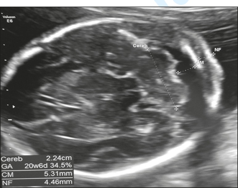
Ultrasound in the median sagittal plane, the best plane in which to assess the fetal posterior fossa (mainly for the differential diagnosis of cystic malformations). It is possible to identify the following: the brainstem (BS); the cerebellar vermis (V), with assessment of its morphology and biometry (craniocaudal and anteroposterior diameter); the primary fissure (yellow arrow); the fourth ventricle; the fastigium (white arrow); and the tentorium (T).

3D ultrasonography of the posterior fossa at 28 weeks of gestation, which allows multiplanar evaluation (OmniView). A: Axial acquisition in the transcerebellar plane. B: Reconstruction in the sagittal plane (OmniView) of the posterior fossa.

Flowchart for the differential diagnosis of posterior fossa anomalies.

Axial ultrasound of the transcerebellar plane, showing communication between the fourth ventricle and the cisterna magna (arrow).

Axial ultrasound of a Dandy-Walker malformation, showing communication between the fourth ventricle and the cisterna magna (white arrow). Sagittal ultrasound showing an abnormal fastigium, vermian agenesis/hypoplasia with upward rotation, and elevation of the tentorium (yellow arrow).

Axial ultrasound of a fetus with vermian hypoplasia, showing communication between the fourth ventricle and the cisterna magna (white arrow). Sagittal ultrasound showing a small vermis, an abnormal fastigium, and the tentorium in a normal position (yellow arrow).

Ultrasound of a fetus with cerebellar hypoplasia, showing a transcerebellar diameter below the 10th percentile for gestational age. The cisterna magna may falsely appear to be increased because the cerebellum is small.

Coronal ultrasound of a fetus with rhombencephalosynapsis, showing that the foliations of the cerebellar hemispheres are continuous in the middle portion of the cerebellum, and that there is no observable vermis (arrowheads). Axial ultrasound can show a cerebellum with a transverse diameter that is normal or below the 10th percentile for gestational age and with a triangular shape.
References
-
- Pilu G, Malinger G, Buyukkurt S. Anomalies of the cerebellum. In: Timor-Tritsch IE, Monteagudo A, Pilu G, et al., editors. Ultrassonography of the prenatal brain. 3rd ed. McGrawHill; 2012. pp. 283–302.
-
- Malinger G, Lev D, Lerman-Sagie T. The fetal cerebellum. Pitfalls in diagnosis and management. Prenat Diagn. 2009;29:372–380. - PubMed
-
- Gandolfi Colleoni G, Contro E, Carletti A, et al. Prenatal diagnosis and outcome of fetal posterior fossa fluid collections. Ultrasound Obstet Gynecol. 2012;39:625–631. - PubMed
-
- Shekdar K. Posterior fossa malformations. Semin Ultrasound CT MR. 2011;32:228–241. - PubMed
-
- Kollias SS, Ball Jr WS, Prenger EC. Cystic malformation of the posterior fossa: differential diagnosis clarified through embryologic analysis. Radiographics. 1993;13:1221–1231. - PubMed
Publication types
LinkOut - more resources
Full Text Sources