. 2019 Nov-Dec;52(6):397-402.
doi: 10.1590/0100-3984.2018.0079.

Twist and shout: magnetic resonance imaging findings in ovarian torsion

Affiliations

Twist and shout: magnetic resonance imaging findings in ovarian torsion

Matheus Menezes Gomes et al. Radiol Bras. 2019 Nov-Dec.

Abstract

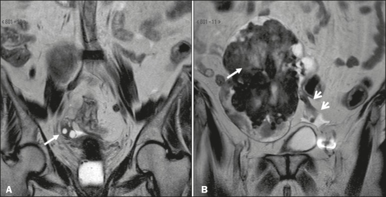
Adnexal torsion is characterized by partial or complete rotation of the suspensory ligament of the ovary and its corresponding vascular pedicle, resulting in vascular impairment that can culminate in hemorrhagic infarction, as well as necrosis of the ovary and fallopian tube. Because there are myriad causes of acute pelvic pain, the differential diagnosis of ovarian torsion is often challenging. Consequently, radiologists should be familiar with the main imaging findings. In this regard, there are typical signs of ovarian torsion on magnetic resonance imaging, including increased ovarian volume with stromal edema and peripheral distribution of the ovarian follicles, as well as thickening of the fallopian tube, an adnexal mass (causal factor) that shifts toward the midline, and the classic, pathognomonic "whirlpool sign". The objective of this essay was to review and illustrate the various magnetic resonance imaging findings in ovarian torsion.

Keywords: Magnetic resonance imaging; Ovarian cysts; Ovarian neoplasms; Ovary; Torsion abnormality.


Figures

Figure 1
Figure 1
Sagittal and coronal T2-weighted MRI sequences (A and B, respectively). A: Preserved ovarian volume and an adjacent hyperintense expansile cystic formation consistent with a paraovarian cyst (arrows). B: Image of another patient with an enlarged ovary due to expansile, hypointense, heterogeneous fibroma-like formation (arrow). Note also the edema at the periphery of the ovarian parenchyma (asterisks).
Figure 2
Figure 2
Coronal T2-weighted MRI sequences. A: Right ovary in a normal position with normal volume (arrow). B: Enlarged left ovary shifted to the right due to the presence of hypointense, heterogeneous expansile formation consistent with a fibroma (long arrow). Note also the ectatic vascular pedicle (short arrows).
Figure 3
Figure 3
Axial T2-weighted MRI sequences. A: Right ovary in a normal position with normal volume (arrow). B: Enlarged left ovary shifted to the right due to the presence of hypointense, heterogeneous expansile formation consistent with a fibroma or fibrothecoma (asterisk). C: Surgical specimen showing a lobulated lesion that proved to be a fibrothecoma.
Figure 4
Figure 4
Coronal and sagittal T2- weighted MRI sequences (A and B, respectively). A: Large adnexal cystic formation (paraovarian cyst, asterisk). Note the hyperintense area, consistent with stromal edema, in the center of the ovary (black arrow), with consequent displacement of follicles to the periphery (white arrow). B: Another large adnexal cystic formation (paraovarian cyst, asterisk) . Again, there is a hyperintense area, consistent with stromal edema, in the center of the ovary (black arrow), with consequent displacement of follicles to the periphery (white arrow).
Figure 5
Figure 5
A–C: Sagittal fat-suppressed T2-weighted SPIR MRI sequences. Note the voluminous mass within the left ovary (asterisks), resulting in adnexal torsion, characterized by the whirlpool sign, with associated flow voids (long arrows), indicative of vascular torsion. The affected ovary (asterisk) is elevated and shows peripheral edema (short arrows). D: Intraoperative image showing the twisted, slightly ischemic adnexal pedicle (dashed outline) and the ovarian lesion that was later identified as a fibrothecoma.
Figure 6
Figure 6
Sagittal T2-weighted MRI sequences, with magnified images below. Note that the ovarian pedicle is twisted around its own axis. The left ovary is enlarged due to the presence of a hypointense, heterogeneous expansile formation consistent with a fibroma (asterisks).
Figure 7
Figure 7
Coronal T2-weighted MRI sequences, with magnified images below. Note that the ovarian pedicle is twisted around its own axis. The left ovary is enlarged due to the presence of a hypointense, heterogeneous expansile formation consistent with a fibroma (asterisks).
Figure 8
Figure 8
Sagittal T2-weighted MRI sequence. Thickened fallopian tube, measuring 1.4 cm in diameter (arrow). Note the large adnexal cystic formation (paraovarian cyst, asterisk).

References

    1. Chang HC, Bhatt S, Dogra VS. Pearls and pitfalls in diagnosis of ovarian torsion. Radiographics. 2008;28:1355–1368. - PubMed
    1. Hibbard LT. Adnexal torsion. Am J Obstet Gynecol. 1985;152:456–461. - PubMed
    1. Huang C, Hong MK, Ding DC. A review of ovary torsion. Ci Ji Yi Xue Za Zhi. 2017;29:143–147. - PMC - PubMed
    1. Breech LL, Hillard PJA. Adnexal torsion in pediatric and adolescent girls. Curr Opin Obstet Gynecol. 2005;17:483–489. - PubMed
    1. Oelsner G, Shashar D. Adnexal torsion. Clin Obstet Gynecol. 2006;49:459–463. - PubMed

LinkOut - more resources