

The microRNA212 regulated PEA15 promotes ovarian cancer progression by inhibiting of apoptosis
- PMID: 32047549
- PMCID: PMC6995389
- DOI: 10.7150/jca.32886
The microRNA212 regulated PEA15 promotes ovarian cancer progression by inhibiting of apoptosis
Abstract
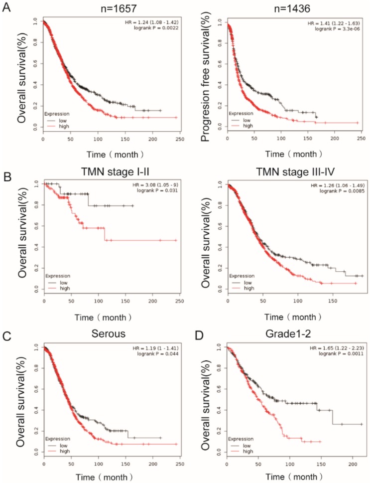
PEA15 (Proliferation And Apoptosis Adaptor) is a 15kDa multifunctional phosphoprotein involved in various essential biological processes such as proliferation and apoptosis of cancer cells. Previous studies have demonstrated that PEA15 can promote the progression of many malignancies. In the present study, the expression of PEA15 in ovarian cancer and normal tissues analyzed in several databases and PEA15 was found to be significantly up-regulated in OC tissues compared to normal tissues. Immunochemical assays performed using 171 OC tissue specimens proved that the expression of PEA15 was remarkably positively correlated with the FIGO stage and associated with histologic subgroups of ovarian cancer. IHC assay for the two phosphorylation sites of PEA15 S116 and S104 was also performed. PEA15 high expression predicted a poor prognosis in OC patients analysed from K-M plot dataset. In addition, we proved knockdown of PEA15 inhibits OC cell proliferation and induces cell apoptosis by Bcl2 downregulation and Bax and cleaved Caspase-3 upregulation. Overexpression of PEA15 promotes the proliferative capacity of OC cells. Moreover, this study first discovered PEA15 expression in OC can be negatively regulated by microRNA212. Overexpression of miR-212 in ovarian cancer cells could cause downregulated the expression of PEA15 expression. Overexpression of miR-212 was found to exerted similar effects on the proliferation, and apoptosis of the ovarian cancer cells as that of PEA15 suppression. Additionally, overexpression of PEA15could at least partially abolished the effects of miR-212 on the proliferation, and apoptosis of ovarian cancer cells. In conclusion, our findings revealed PEA15 appears as a novel predictive biomarker, thus providing a valuable therapeutic target in OC treatment strategy.
Keywords: Apoptosis; OC; PEA15; Proliferation; miR212.
© The author(s).
Conflict of interest statement
Competing Interests: The authors have declared that no competing interest exists.
Figures


Similar articles
-
F-Box and Leucine-Rich Repeat Protein 20 (FBXL20), Negatively Regulated by microRNA (miR)-195-5p, Accelerates the Malignant Progression of Ovarian Cancer.Mol Biotechnol. 2021 Dec;63(12):1235-1243. doi: 10.1007/s12033-021-00375-y. Epub 2021 Aug 2. Mol Biotechnol. 2021. PMID: 34338995
-
Long non-coding RNA PCAT-1 promotes tumor progression by inhibiting miR-129-5p in human ovarian cancer.Arch Med Sci. 2019 Mar;15(2):513-521. doi: 10.5114/aoms.2018.75534. Epub 2018 Apr 30. Arch Med Sci. 2019. PMID: 30899305 Free PMC article.
-
NEAT1 regulates cell proliferation and apoptosis of ovarian cancer by miR-34a-5p/BCL2.Onco Targets Ther. 2017 Oct 6;10:4905-4915. doi: 10.2147/OTT.S142446. eCollection 2017. Onco Targets Ther. 2017. PMID: 29062236 Free PMC article.
-
The protein phosphatase 4 - PEA15 axis regulates the survival of breast cancer cells.Cell Signal. 2016 Sep;28(9):1389-1400. doi: 10.1016/j.cellsig.2016.06.011. Epub 2016 Jun 15. Cell Signal. 2016. PMID: 27317964
-
MicroRNA-506-3p inhibits proliferation and promotes apoptosis in ovarian cancer cell via targeting SIRT1/AKT/FOXO3a signaling pathway.Neoplasma. 2020 Mar;67(2):344-353. doi: 10.4149/neo_2020_190517N441. Epub 2020 Jan 21. Neoplasma. 2020. PMID: 31973537
Cited by
-
Identification of a Novel Protein-Based Signature to Improve Prognosis Prediction in Renal Clear Cell Carcinoma.Front Mol Biosci. 2021 Mar 25;8:623120. doi: 10.3389/fmolb.2021.623120. eCollection 2021. Front Mol Biosci. 2021. PMID: 33842538 Free PMC article.
-
ZNF703 promotes tumor progression in ovarian cancer by interacting with HE4 and epigenetically regulating PEA15.J Exp Clin Cancer Res. 2020 Nov 27;39(1):264. doi: 10.1186/s13046-020-01770-0. J Exp Clin Cancer Res. 2020. PMID: 33246486 Free PMC article.
-
High Expression of PEA15 Is Associated With Patient Survival in Malignant Pleural Mesothelioma.Cancer Diagn Progn. 2021 Jul 3;1(4):371-377. doi: 10.21873/cdp.10049. eCollection 2021 Sep-Oct. Cancer Diagn Progn. 2021. PMID: 35403140 Free PMC article.
-
Identification and verification of a prognostic autophagy-related gene signature in hepatocellular carcinoma.Sci Rep. 2024 Feb 6;14(1):3032. doi: 10.1038/s41598-024-53565-4. Sci Rep. 2024. PMID: 38321105 Free PMC article.
-
Deregulated miRNA clusters in ovarian cancer: Imperative implications in personalized medicine.Genes Dis. 2022 Mar 1;9(6):1443-1465. doi: 10.1016/j.gendis.2021.12.026. eCollection 2022 Nov. Genes Dis. 2022. PMID: 36157483 Free PMC article. Review.
References
-
- La Vecchia C. Ovarian cancer: epidemiology and risk factors. European journal of cancer prevention: the official journal of the European Cancer Prevention Organisation (ECP) 2017;26(1):55–62. - PubMed
-
- Kaldawy A, Segev Y, Lavie O. et al. Low-grade serous ovarian cancer: A review. Gynecologic oncology. 2016;143(2):433–8. - PubMed
-
- Smith RA, Andrews KS, Brooks D. et al. Cancer screening in the United States, 2018: A review of current American Cancer Society guidelines and current issues in cancer screening. CA: a cancer journal for clinicians. 2018;68(4):297–316. - PubMed
LinkOut - more resources
Full Text Sources
Research Materials
