Review
. 2020 Feb 11:6:6.
doi: 10.1038/s41421-020-0141-7. eCollection 2020.

Lysosome biology in autophagy

Affiliations
Review

Lysosome biology in autophagy

Willa Wen-You Yim et al. Cell Discov. .

Abstract

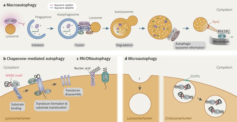
Autophagy is a major intracellular degradation system that derives its degradative abilities from the lysosome. The most well-studied form of autophagy is macroautophagy, which delivers cytoplasmic material to lysosomes via the double-membraned autophagosome. Other forms of autophagy, namely chaperone-mediated autophagy and microautophagy, occur directly on the lysosome. Besides providing the means for degradation, lysosomes are also involved in autophagy regulation and can become substrates of autophagy when damaged. During autophagy, they exhibit notable changes, including increased acidification, enhanced enzymatic activity, and perinuclear localization. Despite their importance to autophagy, details on autophagy-specific regulation of lysosomes remain relatively scarce. This review aims to provide a summary of current understanding on the behaviour of lysosomes during autophagy and outline unexplored areas of autophagy-specific lysosome research.

Keywords: Autophagy; Lysosomes.


Conflict of interest statement

Conflict of interestThe authors declare that they have no conflict of interest.

Figures

Fig. 1
Fig. 1. Autophagy processes.
a Macroautophagy is the only autophagy process that involves another organelle, the autophagosome. It is induced when mTORC1 becomes inactivated upon dissociation from the lysosome. After the phagophore matures into a double-membraned autophagosome, the lysosome fuses with the outer autophagosomal membrane in a SNARE-dependent manner. Fusion is facilitated by tethering factors that bind to proteins on the autophagosome (e.g. LC3) and the lysosome (e.g. RAB7). Lysosomal enzymes degrade the inner autophagosomal membrane and sequestered material. Tubules extend from autolysosomes by KIF5B binding to clathrin-organised PI(4,5)P2 clusters on the autolysosomal membrane and moving away from the autolysosome on microtubules. The tubules are eventually cleaved from the autolysosome by Dyn2, generating new lysosomes. b Chaperone-mediated autophagy (CMA) involves the direct uptake of proteins with the KFERQ(-like) motif into lysosomes via a translocation complex consisting of LAMP2A monomers on the lysosomal membrane that is stabilised by GFAP and luminal HSP90. CMA substrates are delivered to LAMP2A by cytosolic HSC70 and other cytosolic chaperones. Substrate translocation is assisted by lysosomal HSC70. c RN/DNautophagy is the direct delivery of nucleic acids into lysosomes via the nucleic acid transporter, SIDT2. LAMP2C binds nucleic acids and potentially passes them to SIDT2 for translocation into the lysosomal lumen. d Microautophagy is the uptake of cytosolic material by invagination of the lysosomal membrane. Although it has been observed in lysosomes since the discovery of this organelle, mechanistic details are still scarce. Microautophagy in endosomes is more well-understood. Endosomal microautophagy substrates contain KFERQ(-like) motifs and are recognised by cytosolic HSC70 to be delivered to endosomes, where HSC70 binds to phosphatidylserine. Membrane deformation and eventually scission of intralumenal vesicle from the endosomal membrane are executed by the ESCRT machinery.
Fig. 2
Fig. 2. Mechanisms of autophagy machinery recruitment to damaged lysosomes.
Extensive damage to the lysosomal membrane allows cytosolic proteins to pass through freely, including glycan-binding galectins and ubiquitin ligases. Damaged lysosomes are heavily ubiquitinated, which is carried out by ubiquitylation enzymes such as UBE2QL1 (an E2 enzyme), TRIM16 (an E3 ligase) and SCFFBOX27 (an E3 ligase). K48-linked ubiquitin chains are removed by the p97-YOD1-UBXD1-PLAA complex to emphasise the presence of K63-linked chains, which are preferred by the autophagy machinery. Autophagy adaptors bind either directly to galectins (e.g. NDP52) or to ubiquitin (e.g. p62, OPTN, TAX1BP1). They then recruit the autophagy machinery, including the initiation complex, and serve to promote the formation of the autophagosome specifically around the damaged lysosome.

Similar articles

Cited by

References

    1. Mizushima N, Levine B, Cuervo AM, Klionsky DJ. Autophagy fights disease through cellular self-digestion. Nature. 2008;451:1069–1075. doi: 10.1038/nature06639. - DOI - PMC - PubMed
    1. Schroder BA, Wrocklage C, Hasilik A, Saftig P. The proteome of lysosomes. Proteomics. 2010;10:4053–4076. doi: 10.1002/pmic.201000196. - DOI - PubMed
    1. Klionsky DJ, Eskelinen EL. The vacuole versus the lysosome: when size matters. Autophagy. 2014;10:185–187. doi: 10.4161/auto.27367. - DOI - PMC - PubMed
    1. Lawrence RE, Zoncu R. The lysosome as a cellular centre for signalling, metabolism and quality control. Nat. Cell Biol. 2019;21:133–142. doi: 10.1038/s41556-018-0244-7. - DOI - PubMed
    1. Sardiello M, et al. A gene network regulating lysosomal biogenesis and function. Science. 2009;325:473–477. doi: 10.1126/science.1174447. - DOI - PubMed