. 2020 Jan 28:7:9.
doi: 10.3389/fmolb.2020.00009. eCollection 2020.

NMR Methods for Structural Characterization of Protein-Protein Complexes

Affiliations

NMR Methods for Structural Characterization of Protein-Protein Complexes

Jeffrey A Purslow et al. Front Mol Biosci. .

Abstract

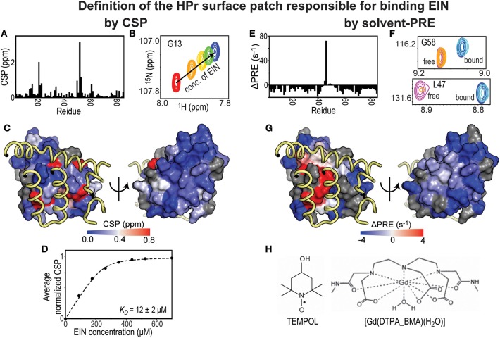
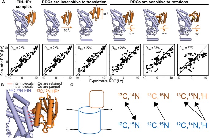
Protein-protein interactions and the complexes thus formed are critical elements in a wide variety of cellular events that require an atomic-level description to understand them in detail. Such complexes typically constitute challenging systems to characterize and drive the development of innovative biophysical methods. NMR spectroscopy techniques can be applied to extract atomic resolution information on the binding interfaces, intermolecular affinity, and binding-induced conformational changes in protein-protein complexes formed in solution, in the cell membrane, and in large macromolecular assemblies. Here we discuss experimental techniques for the characterization of protein-protein complexes in both solution NMR and solid-state NMR spectroscopy. The approaches include solvent paramagnetic relaxation enhancement and chemical shift perturbations (CSPs) for the identification of binding interfaces, and the application of intermolecular nuclear Overhauser effect spectroscopy and residual dipolar couplings to obtain structural constraints of protein-protein complexes in solution. Complementary methods in solid-state NMR are described, with emphasis on the versatility provided by heteronuclear dipolar recoupling to extract intermolecular constraints in differentially labeled protein complexes. The methods described are of particular relevance to the analysis of membrane proteins, such as those involved in signal transduction pathways, since they can potentially be characterized by both solution and solid-state NMR techniques, and thus outline key developments in this frontier of structural biology.

Keywords: chemical shift perturbations; isotopic labeling; residual dipolar couplings; solid state NMR; solvent-PRE.


Figures

Figure 1
Figure 1
Solvent-PRE and CSP analysis of EIN-HPr complex. (A) CSP measured for 15N-labeled HPr in the presence of saturating concentrations of unlabeled EIN are plotted vs. residue index. (B) Example cross-peak from 1H-15N HSQC spectrum of 15N-labeled HPr measured at increasing concentration of EIN. A peak from the complex interface was selected. (C) The CSP data from panel (A) are plotted on the surface of HPr according to the color bar. The relevant portions of EIN are shown as yellow tubes. (D) CSP vs. concertation of EIN (black circles). The data can be fit (black line) to return the KD of the EIN-HPr complex. (E) ΔPRE vs. residue index. ΔPREs are calculated by subtracting the solvent-PREs (i.e., the increase in 1HN-R2 caused by addition of 4 mM Gd(DTPA-BMA) to the NMR sample) measured for 15N-labeled HPr complexed to unlabeled EIN from the solvent-PRE data measured for the free protein. While the majority of the HPr residues show a negative ΔPRE (which is the result of the reduced rotational diffusion of complexed HPr compared to the free protein), obstruction of the paramagnetic probe from the binding interface results in positive ΔPREs. (F) Example cross-peaks from 1H-15N HSQC spectra of 0.8 mM 15N-labeled HPr in the presence of 0 mM EIN and 0 mM Gd(DTPA-BMA) (pink), 0 mM EIN and 4 mM Gd(DTPA-BMA) (orange), 1 mM EIN and 0 mM Gd(DTPA-BMA) (blue), 1 mM EIN and 4 mM Gd(DTPA-BMA) (cyan). The two cross-peaks have been chosen to illustrate the cases of a residue located far from the complex interface (G58) and of an HPr residue that is in direct contact with EIN (L47). (G) ΔPREs are plotted on the surface of HPr according to the color bar. The relevant portions of EIN are shown as yellow tubes. (H) Structures of two commonly used paramagnetic probes for surface accessibility studies.
Figure 2
Figure 2
NMR methods for structure determination of macromolecular complexes. (A) Fitting of the experimental N-HN RDCs measured for the phosphorylated EIN-HPr complex (Suh et al., 2008) to (i) the experimental NMR structure (PDB code: 3EZA; left panel), (ii) two structural models generated by 10 Å translation of HPr along two perpendicular directions (seconds and third panels from left), and (iii) three structural models generated by rotations of 10°, 25°, and 45° of HPr about an axis perpendicular to the complex interface (last three panels from left). The quality of the fit is judged in terms of R-factor. A high R-factor indicates poor agreement between the experimental RDC data and the structural model (Clore and Garrett, 1999). (B) In an isotope-edited/isotope-filtered experiment intramolecular NOEs (black dashed lines) are purged, while intermolecular NOEs (red solid lines) are retained. In panels (A,B) HPr is orange and EIN is blue. (C) Schematic representation of a solid protein complex and heteronuclear polarization transfer schemes. In the first scheme, mixtures of isotopic labeling allow intermolecular heteronuclear correlations. In the seconds scheme, intramolecular 15N-13C dephasing precedes intermolecular heteronuclear 15N-13C correlations. In the third scheme, intramolecular 1H11H-13C33C/15N55N dephasing facilitates intermolecular 1H-13C/15N cross-polarization that identifies isotopically labeled sites on a protein interacting with an unlabeled binding partner.

References

    1. Anglister J., Srivastava G., Naider F. (2016). Detection of intermolecular NOE interactions in large protein complexes. Prog. Nucl. Magn. Reson. Spectrosc. 97, 40–56. 10.1016/j.pnmrs.2016.08.002 - DOI - PubMed
    1. Anthis N. J., Clore G. M. (2015). Visualizing transient dark states by NMR spectroscopy. Q. Rev. Biophys. 48, 35–116 10.1017/S0033583514000122 - DOI - PMC - PubMed
    1. Arachchige R. J., Burton S. D., Lu J.-X., Ginovska B., Harding L. K., Taylor M. E., et al. (2018). Solid-state NMR identification of intermolecular interactions in amelogenin bound to hydroxyapatite. Biophys. J. 115, 1666–1672. 10.1016/j.bpj.2018.08.027 - DOI - PMC - PubMed
    1. Arumugam S., Hemme C. L., Yoshida N., Suzuki K., Nagase H., Berjanskii M., et al. (1998). TIMP-1 contact sites and perturbations of stromelysin 1 mapped by NMR and a paramagnetic surface probe. Biochemistry 37, 9650–9657. 10.1021/bi980128h - DOI - PubMed
    1. Bayro M. J., Debelouchina G. T., Eddy M. T., Birkett N. R., MacPhee C. E., Rosay M., et al. (2011). Intermolecular structure determination of amyloid fibrils with magic-angle spinning and dynamic nuclear polarization NMR. J. Am. Chem. Soc. 133, 13967–13974. 10.1021/ja203756x - DOI - PMC - PubMed

LinkOut - more resources