

Spaceflight associated neuro-ocular syndrome (SANS) and the neuro-ophthalmologic effects of microgravity: a review and an update
- PMID: 32047839
- PMCID: PMC7005826
- DOI: 10.1038/s41526-020-0097-9
Spaceflight associated neuro-ocular syndrome (SANS) and the neuro-ophthalmologic effects of microgravity: a review and an update
Erratum in
-
Erratum: Author Correction: Spaceflight associated neuro-ocular syndrome (SANS) and the neuro-ophthalmologic effects of microgravity: a review and an update.NPJ Microgravity. 2020 Aug 26;6:23. doi: 10.1038/s41526-020-00114-8. eCollection 2020. NPJ Microgravity. 2020. PMID: 32885041 Free PMC article.
Abstract
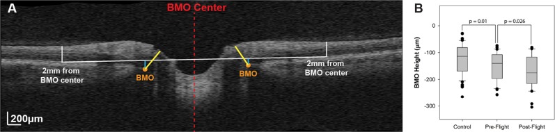
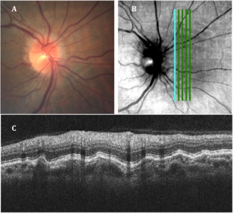
Prolonged microgravity exposure during long-duration spaceflight (LDSF) produces unusual physiologic and pathologic neuro-ophthalmic findings in astronauts. These microgravity associated findings collectively define the "Spaceflight Associated Neuro-ocular Syndrome" (SANS). We compare and contrast prior published work on SANS by the National Aeronautics and Space Administration's (NASA) Space Medicine Operations Division with retrospective and prospective studies from other research groups. In this manuscript, we update and review the clinical manifestations of SANS including: unilateral and bilateral optic disc edema, globe flattening, choroidal and retinal folds, hyperopic refractive error shifts, and focal areas of ischemic retina (i.e., cotton wool spots). We also discuss the knowledge gaps for in-flight and terrestrial human research including potential countermeasures for future study. We recommend that NASA and its research partners continue to study SANS in preparation for future longer duration manned space missions.
Keywords: Eye manifestations; Medical research; Physical examination.
© The Author(s) 2020.
Conflict of interest statement
Competing interestsAll of the authors declare that they have no “competing interests” related to funding, person, or financial interest. Although the authors work directly (W.T., T.B.) as employees or indirectly as consultants (A.G.L., C.R.G.) for NASA, the views and opinions expressed here are those of the authors and do not necessarily reflect the views of NASA or the United States government. Dr. Lee has served as treating physician and expert witness but no cases involved Space Flight associated Neuro-ocular syndrome (SANS) or any of the content of this paper. In addition, the remaining authors do not have any of the following non-financial interests: Unpaid membership in a government or non-governmental organization; unpaid membership in an advocacy or lobbying organization; unpaid advisory position in a commercial organization; Writing or consulting for an educational company; or Acting as an expert witness.
Figures

References
Publication types
LinkOut - more resources
Full Text Sources
Other Literature Sources
