Case Reports
. 2020 May;139(5):875-892.
doi: 10.1007/s00401-020-02132-y. Epub 2020 Feb 11.

The pathology of central nervous system inflammatory demyelinating disease accompanying myelin oligodendrocyte glycoprotein autoantibody

Affiliations
Case Reports

The pathology of central nervous system inflammatory demyelinating disease accompanying myelin oligodendrocyte glycoprotein autoantibody

Romana Höftberger et al. Acta Neuropathol. 2020 May.

Abstract

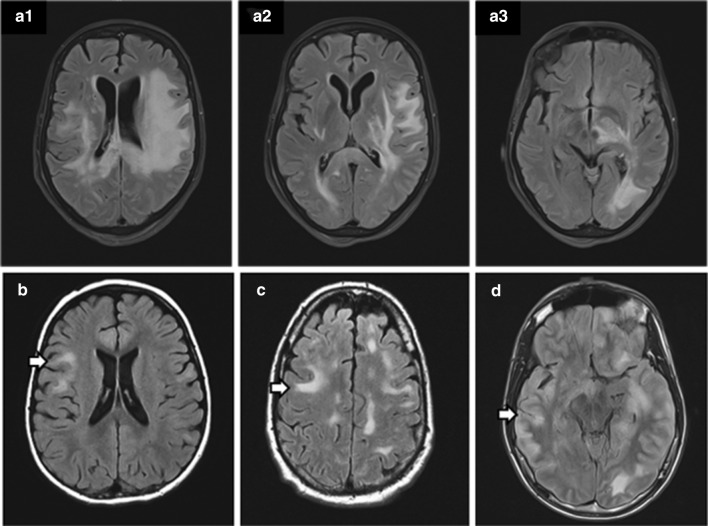
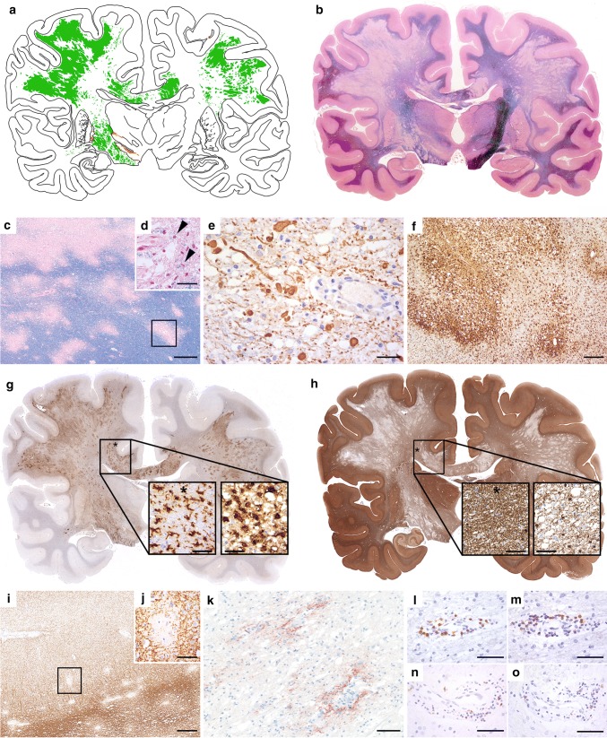
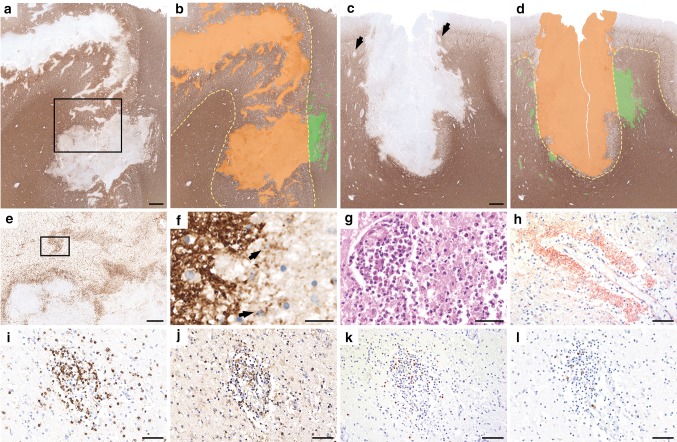
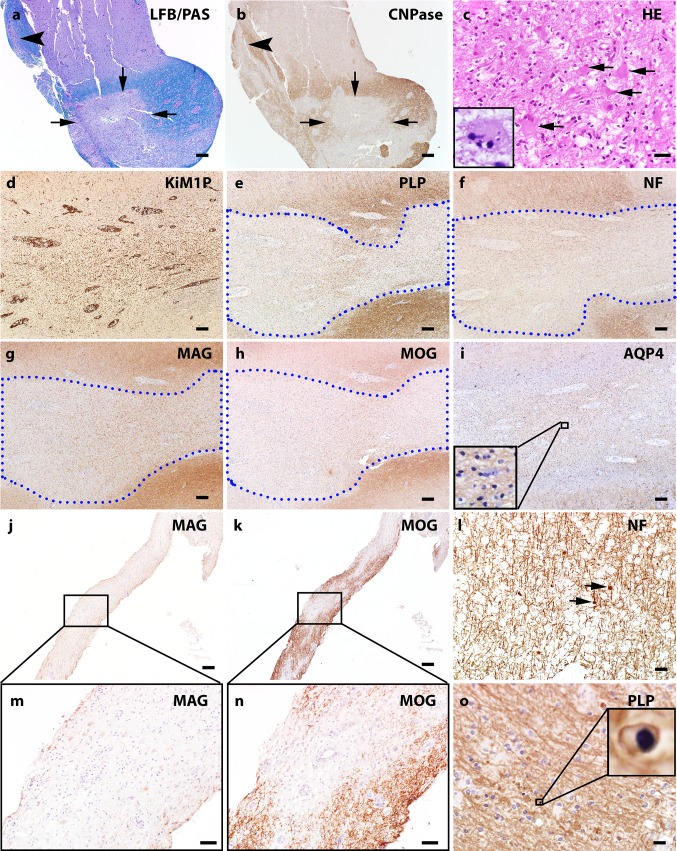
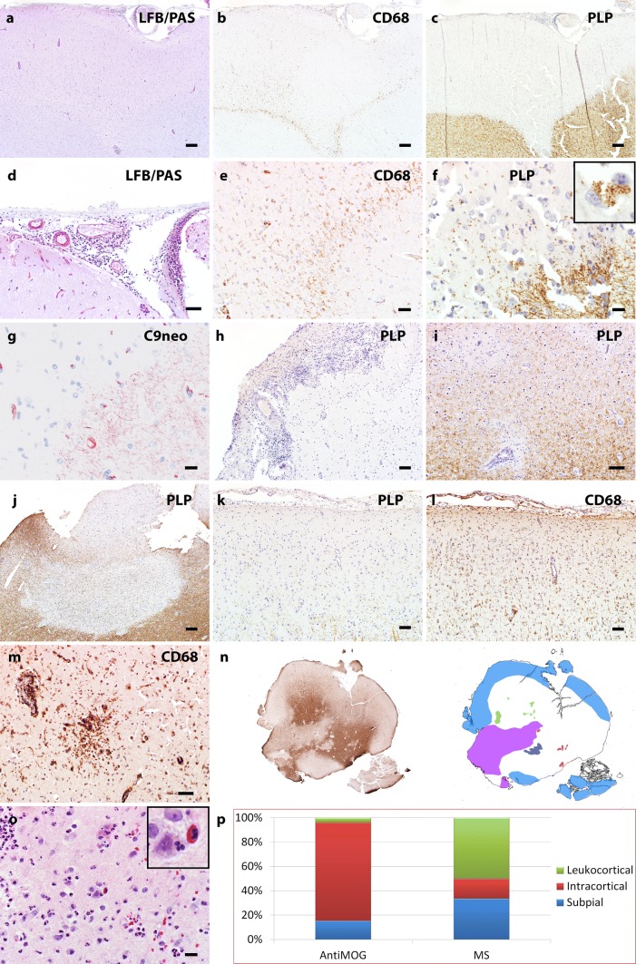
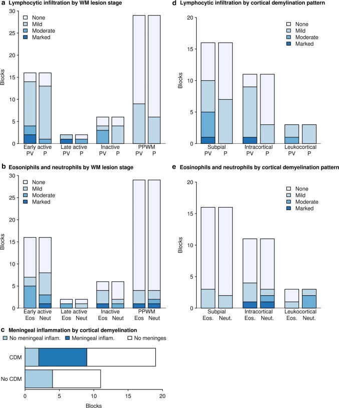
We sought to define the pathological features of myelin oligodendrocyte glycoprotein (MOG) antibody associated disorders (MOGAD) in an archival autopsy/biopsy cohort. We histopathologically analyzed 2 autopsies and 22 brain biopsies from patients with CNS inflammatory demyelinating diseases seropositive for MOG-antibody by live-cell-based-assay with full length MOG in its conformational form. MOGAD autopsies (ages 52 and 67) demonstrate the full spectrum of histopathological features observed within the 22 brain biopsies (median age, 10 years; range, 1-66; 56% female). Clinical, radiologic, and laboratory characteristics and course (78% relapsing) are consistent with MOGAD. MOGAD pathology is dominated by coexistence of both perivenous and confluent white matter demyelination, with an over-representation of intracortical demyelinated lesions compared to typical MS. Radially expanding confluent slowly expanding smoldering lesions in the white matter as seen in MS, are not present. A CD4+ T-cell dominated inflammatory reaction with granulocytic infiltration predominates. Complement deposition is present in all active white matter lesions, but a preferential loss of MOG is not observed. AQP4 is preserved, with absence of dystrophic astrocytes, and variable oligodendrocyte and axonal destruction. MOGAD is pathologically distinguished from AQP4-IgG seropositive NMOSD, but shares some overlapping features with both MS and ADEM, suggesting a transitional pathology. Complement deposition in the absence of selective MOG protein loss suggest humoral mechanisms are involved, however argue against endocytic internalization of the MOG antigen. Parallels with MOG-EAE suggest MOG may be an amplification factor that augments CNS demyelination, possibly via complement mediated destruction of myelin or ADCC phagocytosis.

Keywords: Acute disseminated encephalomyelitis; Autopsy; Biopsy; Demyelination; MOG; Multiple sclerosis.


Conflict of interest statement

No authors had conflicts of interests relevant to this study.

Figures

Fig. 1
Fig. 1
MRI findings in patients with MOG-associated encephalomyelitis. a1–a3 Brain MRI from autopsy patient 1, obtained on day 40 after symptom onset, shows extensive, confluent T2 abnormalities involving the subcortical white matter of the frontal, temporal, and occipital lobe, bilaterally, with contrast enhancement in one fronto-parietal lesion (not shown) and extension into deep grey matter. b–d MRI of 3 MOGAD biopsied cases just prior to surgery with the arrow reflecting the site of biopsy
Fig. 2
Fig. 2
Neuropathology of autopsy case 1. Topographic evaluation shows prominent bi-hemispheric demyelination in the white matter, focally extending into the cortex and deep grey matter (a, green: white matter demyelination, orange: grey matter demyelination; b, LFB). The demyelinated lesions are partly confluent with massive perivenous accentuation (c, LFB) and characterized by early active demyelination with LFB-positive degradation products within the macrophage cytoplasms (d, arrow heads; LFB). Axons are moderately reduced and form numerous axonal spheroids (e, SMI31). Some of the confluent demyelinating lesions show a transition between perivascular accentuation to a rim of activated macrophages and microglia at the edge (f, HLADR), profound microglia activation is also seen in the periplaque white matter (g, asterisk in PPWM; microglia in PPWM enlarged in left image; macrophages in plaque enlarged in right image; HLADR). Within the demyelinated lesions a higher number of oligodendrocytes is visible compared to NAWM (h, asterisk in NAWM; oligodendrocytes in NAWM enlarged in left image, plaque enlarged in right image; TPPP/p25). Small intracortical perivenous demyelination is also present in the cortex (i, j; rectangle in i enlarged). Within the lesions profound deposition of activated complement complex is visible (k, C9neo antigen). The inflammatory reaction is characterized by perivascular cuffs of moderate numbers of CD3 + (l), CD4 + (m), and CD8 + T cells (n), and only few CD79a + B cells (o). Scale bars c 600 μm; d, h, j 60 μm; i 380 μm; k 100 μm; e, g, l–o 50 μm; f 300 μm
Fig. 3
Fig. 3
Neuropathology of autopsy case 2. Histopathology of autopsy case 2 shows predominantly cortical plaques that extend into the subcortical white matter (a–d, MOG; b and d schematic drawings, cortical demyelination: orange; white matter demyelination green;) and form multiple perivenous areas of demyelination in the adjacent grey and white matter (c, arrows). Activated microglia and macrophages form a rim at the lesion borders and show a perivenous accentuation (e, HLADR; rectangle in a enlarged in e) with MOG-positive demyelination products within the macrophages (f, MOG; arrows; rectangle in e enlarged in f). Some lesions show superimposed ischemic damage with tissue necrosis with massive infiltration of neutrophilic granulocytes (g, H&E). Profound perivenous deposition of activated complement complex is visible (h, C9neo antigen). The inflammatory infiltrates mainly contain CD3 (i) and CD4 positive T cells (j), less CD8-positive T cells (k) and only few perivascular CD79a positive B cells (l). Scale bars a–d 1.2 mm; e 600 μm; f 30 μm; g–l 60 μm
Fig. 4
Fig. 4
White matter pathology of MOG Ab positive inflammatory demyelinating disease. a LFB/PAS stain and myelin protein CNPase immunohistochemistry (b) on consecutive sections indicate the perivenous (arrowhead) and confluent (arrows) demyelination that coexists in the subcortical white matter of a MOGAD biopsy. c H&E stain shows marked hypertrophic reactive astrocytes present in the white matter lesion and Creutzfeldt-Peters cells are occasionally noted (inset). d–i Consecutive sections: d KiM1P immunohistochemistry indicates extensive microglia/macrophage infiltration in the white matter with no obvious border. The blue dotted lines contour the demyelinating lesion e with relative preserved axons f. The loss of minor myelin protein MAG (g) and MOG (h) are equal. AQP4 is preserved in the lesion i. j and k consecutive sections. Preferential MAG loss (j, m) with relative MOG preservation (k, n) is seen in a single MOGAD case. l Mild axonal damage characterized by axonal spheroid (indicated with arrows) is present in the demyelinating lesions. o Apoptotic oligodendrocytes with condensed nucleus (highlighted in the inset) are seen in the lesions. Scale bars in a, b, d–k = 200 μm. Scale bars in c, l and o = 20 μm
Fig. 5
Fig. 5
Cortical pathology of MOG-antibody positive inflammatory demyelinating disease.a–g Consecutive sections. a LFB/PAS staining of the MOGAD cortical biopsy indicates pale cortical staining and meningeal inflammation (the high power image in panel d highlights the meningeal infiltration). b CD68 immunohistochemistry on the consecutive section to a shows microglia/macrophage reactivity in the cortex. The higher power image of b (panel e) highlights microglia/macrophage reactivity at the border of a cortical lesion. c PLP immunohistochemistry indicates extensive cortical myelin loss. f High power image shows demyelinating activity at the lesion border characterized by myelin debris laden macrophages (inset). g C9neo indicates complement deposition on the myelin fibers in the cortex. h PLP immunohistochemistry with hematoxylin counterstain highlights prominent meningeal inflammation associated with extensive subpial cortical demyelination. Focal intracortical (i) and leukocortical (j) lesions are also present in the MOGAD biopsies. In a subpial cortical lesion (k), extensive cortical microglial reactivity is marked (l). Microglial aggregates are occasionally seen in the cortex (m). n PLP staining scan and corresponding schematic figure highlights the complexity of the cortical lesions in a MOGAD case. Light Blue filled square, subpial demyelination. Green filled square, intracortical demyelination; violet filled square, confluent white matter demyelination; red filled square, perivascular white matter demyelination; dark blue filled square, coalescent white matter demyelination. o HE stain highlights eosinophils and neutrophils in the cortex. p The comparison figure shows the different ratios of cortical lesion patterns between MOGAD and multiple sclerosis. Scale bars in a, b, c, h, j, k, l = 200 μm. Scale bars in e, f, g, o = 20 μm. Scale bar in i = 100 μm. Scale bar in m = 50 μm
Fig. 6
Fig. 6
Association between inflammations and demyelination in MOGAD. a The extent of lymphocytic infiltration relative to white matter demyelinating stage for both perivascular (PV) and parenchymal (P) infiltration. Early active and late active demyelinating lesions shows more obvious lymphocytic infiltration in terms of severity and frequency compared to non-demyelinated periplaque white matter (PPWM). b Eosinophil (Eos) and neutrophil (Neut) infiltration is more prominent in white matter lesions compared to the PPWM. c Meningeal infiltration is only present in tissue blocks with cortical demyelination. d Both perivascular and diffuse parenchymal lymphocytic inflammation are present in cortical demyelinating lesions. Perivascular inflammation tends to be more prominent. e Eosinophils and neutrophils are present in all cortical lesion subtypes, but more often present in intracortical lesions

References

    1. Aboul-Enein F, Rauschka H, Kornek B, Stadelmann C, Stefferl A, Bruck W, et al. Preferential loss of myelin-associated glycoprotein reflects hypoxia-like white matter damage in stroke and inflammatory brain diseases. J Neuropathol Exp Neurol. 2003;62:25–33. doi: 10.1093/jnen/62.1.25. - DOI - PubMed
    1. Absinta M, Sati P, Masuzzo F, Nair G, Sethi V, Kolb H, et al. Association of chronic active multiple sclerosis lesions with disability in vivo. JAMA Neurol. 2019 doi: 10.1001/jamaneurol.2019.2399. - DOI - PMC - PubMed
    1. Berger T, Rubner P, Schautzer F, Egg R, Ulmer H, Mayringer I, et al. Antimyelin antibodies as a predictor of clinically definite multiple sclerosis after a first demyelinating event. N Engl J Med. 2003;349:139–145. doi: 10.1056/NEJMoa022328. - DOI - PubMed
    1. Breithaupt C, Schafer B, Pellkofer H, Huber R, Linington C, Jacob U. Demyelinating myelin oligodendrocyte glycoprotein-specific autoantibody response is focused on one dominant conformational epitope region in rodents. J Immunol. 2008;181:1255–1263. doi: 10.4049/jimmunol.181.2.1255. - DOI - PubMed
    1. Bruck W, Porada P, Poser S, Rieckmann P, Hanefeld F, Kretzschmar HA, et al. Monocyte/macrophage differentiation in early multiple sclerosis lesions. Ann Neurol. 1995;38:788–796. doi: 10.1002/ana.410380514. - DOI - PubMed

Publication types

MeSH terms