

Consensus recommendations for a standardized brain tumor imaging protocol for clinical trials in brain metastases
- PMID: 32048719
- PMCID: PMC7283031
- DOI: 10.1093/neuonc/noaa030
Consensus recommendations for a standardized brain tumor imaging protocol for clinical trials in brain metastases
Abstract
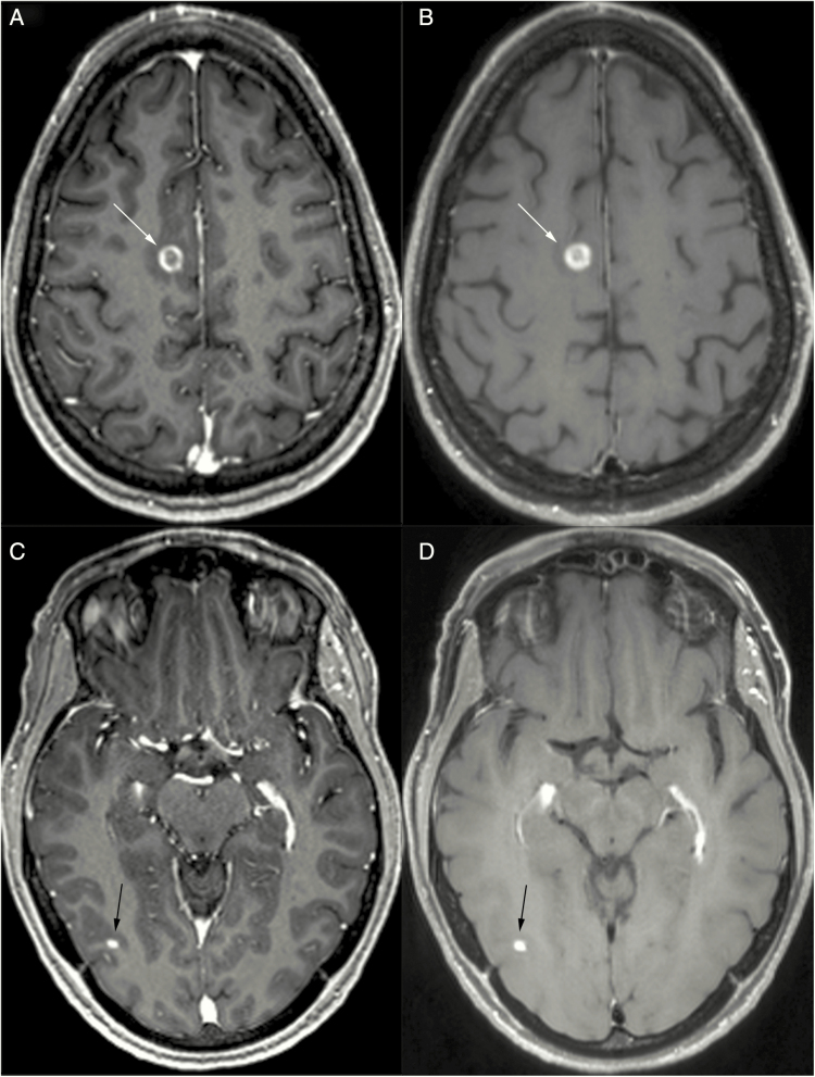
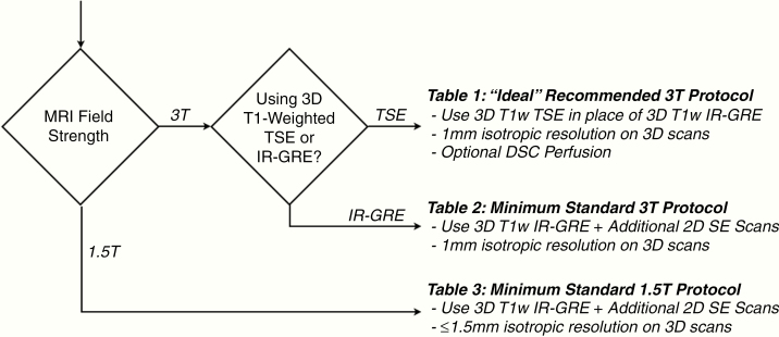
A recent meeting was held on March 22, 2019, among the FDA, clinical scientists, pharmaceutical and biotech companies, clinical trials cooperative groups, and patient advocacy groups to discuss challenges and potential solutions for increasing development of therapeutics for central nervous system metastases. A key issue identified at this meeting was the need for consistent tumor measurement for reliable tumor response assessment, including the first step of standardized image acquisition with an MRI protocol that could be implemented in multicenter studies aimed at testing new therapeutics. This document builds upon previous consensus recommendations for a standardized brain tumor imaging protocol (BTIP) in high-grade gliomas and defines a protocol for brain metastases (BTIP-BM) that addresses unique challenges associated with assessment of CNS metastases. The "minimum standard" recommended pulse sequences include: (i) parameter matched pre- and post-contrast inversion recovery (IR)-prepared, isotropic 3D T1-weighted gradient echo (IR-GRE); (ii) axial 2D T2-weighted turbo spin echo acquired after injection of gadolinium-based contrast agent and before post-contrast 3D T1-weighted images; (iii) axial 2D or 3D T2-weighted fluid attenuated inversion recovery; (iv) axial 2D, 3-directional diffusion-weighted images; and (v) post-contrast 2D T1-weighted spin echo images for increased lesion conspicuity. Recommended sequence parameters are provided for both 1.5T and 3T MR systems. An "ideal" protocol is also provided, which replaces IR-GRE with 3D TSE T1-weighted imaging pre- and post-gadolinium, and is best performed at 3T, for which dynamic susceptibility contrast perfusion is included. Recommended perfusion parameters are given.
Keywords: MRI; brain metastases; imaging; protocol.
© The Author(s) 2020. Published by Oxford University Press on behalf of the Society for Neuro-Oncology. All rights reserved. For permissions, please e-mail: journals.permissions@oup.com.
Figures
Comment in
-
Letter regarding "Consensus recommendations for a standardized brain tumor imaging protocol for clinical trials in brain metastases".Neuro Oncol. 2020 Nov 26;22(11):1705. doi: 10.1093/neuonc/noaa176. Neuro Oncol. 2020. PMID: 32735663 Free PMC article. No abstract available.
-
Response to Letter to Editor.Neuro Oncol. 2020 Nov 26;22(11):1706-1707. doi: 10.1093/neuonc/noaa202. Neuro Oncol. 2020. PMID: 32823280 Free PMC article. No abstract available.
References
-
- Tabouret E, Chinot O, Metellus P, Tallet A, Viens P, Gonçalves A. Recent trends in epidemiology of brain metastases: an overview. Anticancer Res. 2012;32(11):4655–4662. - PubMed
-
- Wen PY, Black PM, Loeffler JS. Metastatic brain cancer. In: DeVita V, Hellman S, Rosenberg SA, eds. Cancer: Principles and Practice of Oncology. 6th ed Philadelphia, PA: Lippincott, Williams, & Wilkins; 2001:2655–2670.
