Review
. 2020 Mar;184(1):7-22.
doi: 10.1002/ajmg.c.31778. Epub 2020 Feb 12.

Development of the human heart

Affiliations
Review

Development of the human heart

Marieke F J Buijtendijk et al. Am J Med Genet C Semin Med Genet. 2020 Mar.

Abstract

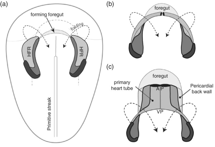
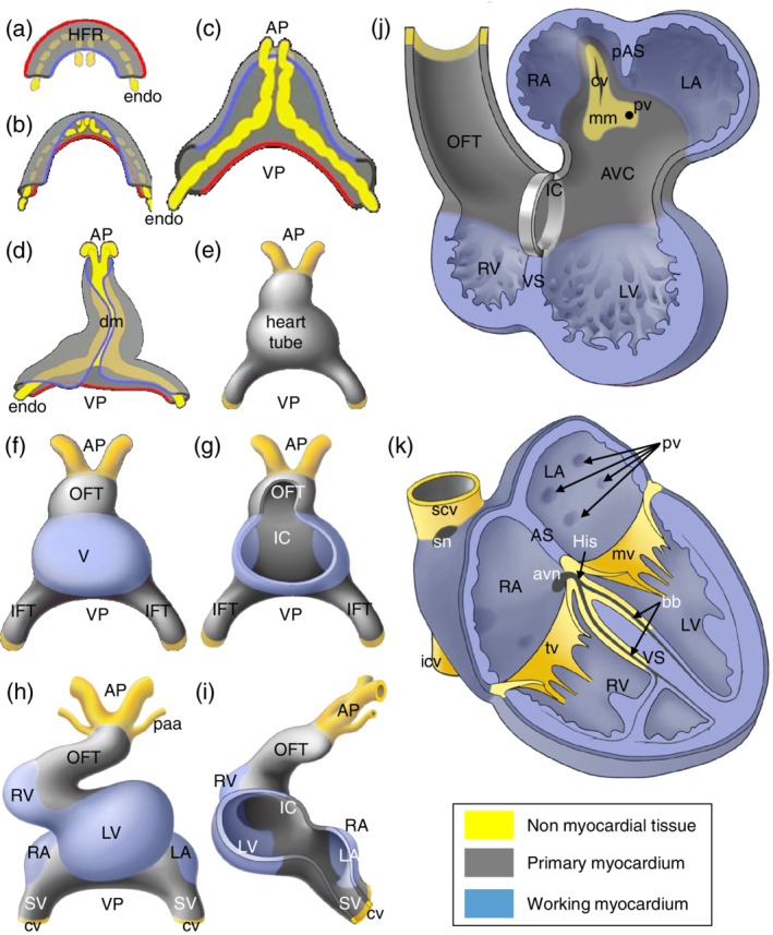
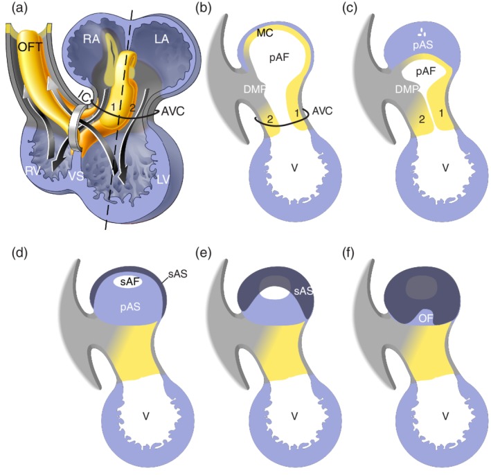
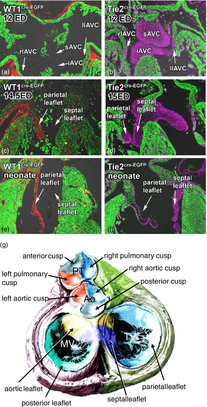
In 2014, an extensive review discussing the major steps of cardiac development focusing on growth, formation of primary and chamber myocardium and the development of the cardiac electrical system, was published. Molecular genetic lineage analyses have since furthered our insight in the developmental origin of the various component parts of the heart, which currently can be unambiguously identified by their unique molecular phenotype. Moreover, genetic, molecular and cell biological analyses have driven insights into the mechanisms underlying the development of the different cardiac components. Here, we build on our previous review and provide an insight into the molecular mechanistic revelations that have forwarded the field of cardiac development. Despite the enormous advances in our knowledge over the last decade, the development of congenital cardiac malformations remains poorly understood. The challenge for the next decade will be to evaluate the different developmental processes using newly developed molecular genetic techniques to further unveil the gene regulatory networks operational during normal and abnormal cardiac development.

Keywords: cardiac development; cardiac growth; epicardial development; septation; valve development.


Figures

Figure 1
Figure 1
From heart forming region to primary heart tube. Panels (a)–(c) provide a schematic representation of the formation of the linear heart tube from the heart forming region (HFR), as seen from the ventral side. Within the HFR the first heart field is indicated in light gray, the second heart field in dark gray, and the third heart field in black. AP refers to the arterial pole and VP to the venous pole of the primary heart tube
Figure 2
Figure 2
Schematic representation of the formation of the adult heart. With the onset of embryonic folding the left and right heart forming region (HFR). The yellow structures represent the endocardial cells (endo) that form the inner lining of the heart and in gray the formed primary myocardium. VP refers to the venous pole were the blood will enter the heart and AP to the arterial pole where the blood will leave the heart. For easy comparison of panels (a) through (d), a red line indicates the lateral border of the HFR and a blue line the medial border. With ongoing folding the HFR becomes positioned ventrally of the foregut (see also Figure 1). At the position where the lateral borders of the HFR meet, the linear heart tube is connected to the body wall via the dorsal mesocardium (dm) (panel d). The DM breaks due to which the heart is only attached to the body wall at the AP and VP (panel h–k). Whereas panels (a)–(d) show a dorsal view of the forming heart, panels (e)–(h) show a ventral view, illustrating the transition of linear heart tube to four‐chambered heart. Only at the ventral site of the linear heart tube the differentiation of the embryonic ventricle (V) is locally initiated. The forming working myocardium of the chambers is indicated in blue (panels f–k). Note in panel (g) that at the dorsal side of the heart tube, primary myocardium is retained, which is referred to as the inner curvature (IC). Flanking the forming ventricle, primary myocardium is retained which is referred to as the inflow tract (IFT) and outflow tract (OFT). With ongoing development, the linear heart tube loops to the right and chamber formation becomes evident (panel h). At this stage the right ventricle (RV) starts to form when primary myocardium of the OFT differentiates into chamber myocardium. Moreover, upstream of the left ventricle (LV), the primary myocardium of the IFT locally differentiates into the left atrium (LA) and right atrium (RA). In the meantime, newly differentiated cardiomyocytes are added to the lengthening heart forming the sinus venous (SV) myocardium. The heart is connected to the blood circulation at the VP via the left and right cardinal vein (cv) and at the AP via the pharyngeal arch arteries (paa). Panel (j) shows a representation of the 5 week old human heart showing the expanding (ballooning) atria and ventricles, as well as the remnants of the primary myocardium of the IFT, AVC (atrioventricular canal), IC and OFT. The forming primary atrial septum (pAS) and ventricular septum (VS) are identified. Within the LA the attachment to the body wall is identified as the mediastinal mesenchyme (mm) through which the cardinal vein (cv) and the future pulmonary vein (pv) drain into the heart. In the formed heart (panel k) the primary myocardium of the IFT and AVC has differentiated into the central conduction system, comprising the sinoatrial node (sn), the atrioventricular node (avn), the His bundle (His) and the bundle branches (bb). Within the right atrium the superior and inferior caval veins (scv and icv) drain in the RA and the pulmonary veins (pv) in the LA. Flanking the chambers, valves are formed of which only the mitral valve (mv) and the tricuspid valve (tv) are shown. The semilunar valves cannot be shown in this representation
Figure 3
Figure 3
Schematic representation of septation. Panel (a) shows the same schematic drawing of the four chamber‐forming heart as Figure 2(j), in which now the major cushions have been added within the atrioventricular canal (AVC) and outflow tract (OFT) and the left and right bloodstreams through the heart is indicated. The ventricular foramen is located between the distal edge of the ventricular septum (VS) and the inner curvature (IC). Noteworthy is that the left bloodstream passes the ventricular foramen during systole, while the right bloodstream already passes through the ventricular foramen during diastole. Panels (b)–(f) illustrate atrial septation and show the cutting edge (sagittal sections) at the level of the dashed line in panel (a). For easy comparison the cushions are numbered. The mesenchymal complex formed by the anterior (1) and posterior (2) atrioventricular cushion, the extension of the anterior cushion over the roof of the atrium (MC), and the extracardiac mesenchyme that protrudes into atrial lumen (DMP), surround the connection between the left (LA) and right atrium (RA). With expansion of this complex the primary atrial foramen (pAF) becomes smaller and eventually closes forming the primary atrial septum (pAS). Prior to closure of the pAF, the secondary atrial foramen (sAS) is formed. Within the RA the secondary atrial septum (sAS) folds down from the atrial wall into the lumen and covers the pAS partly and the pAF completely. The uncovered part of the pAS is recognized as the oval fossa (OF) (Modified from Sylva et al., 2014)
Figure 4
Figure 4
Formation of the valves. Panels (a)–(f) show the formation of the atrioventricular valves. Genetic lineage tracing shows the contribution of the endocardially‐derived (panels b, d, and f) and epicardially‐derived (panels a, c, and e) cells to the cushions and subsequent valve leaflets. The myocardium of the heart is shown in green and the epicardially‐derived cells labeled using the WT1(BAC)‐Cre in red and the endocardially‐derived cells using Tie2‐Cre in magenta (Modified from Wessels et al., 2012). Panel (g) shows a schematic summary of the origin of the cellular contributions to the mitral, tricuspid and semilunar valves. In the pulmonary trunk The left cusp in both the pulmonary trunk and aorta is derived from the septal OFT cushion (blue). The right cusp in both the pulmonary trunk (PT) and aorta (Ao) is derived from the parietal OFT cushion (orange). The anterior cusp in the pulmonary trunk is derived from the right intercalated ridge and in the aorta the posterior cusp is derived from the left intercalated ridge (gray). The aortic or anterior leaflet of the mitral valve (MV) and the septal leaflet of the tricuspid valve (TV) are derived from the inferior (blue) and posterior (yellow) atrioventricular cushions (iAVC and sAVC, respectively). The posterior leaflet of the MV is derived from the left lateral atrioventricular cushion (llAVC, green) and the septal leaflet of the TV from the right lateral atrioventricular cushion (rlAVC, light blue). From the data shown in panels (a)–(f), one can infer that the posterior MV leaflet and the parietal leaflet of the TV comprise predominantly of epicardially‐derived cells and both the aortic leaflet of the MV and the septal leaflet of the TV of endocardially‐derived cells. (Modified from Lamers & Moorman, 2002)

References

    1. Aanhaanen, W. T. , Boukens, B. J. , Sizarov, A. , Wakker, V. , de Gier‐de Vries, C. , van Ginneken, A. C. , … Christoffels, V. M. (2011). Defective Tbx2‐dependent patterning of the atrioventricular canal myocardium causes accessory pathway formation in mice. Journal of Clinical Investigation, 121(2), 534–544. - PMC - PubMed
    1. Aanhaanen, W. T. , Mommersteeg, M. T. , Norden, J. , Wakker, V. , de Gier‐de Vries, C. , Anderson, R. H. , … Christoffels, V. M. (2010). Developmental origin, growth, and three‐dimensional architecture of the atrioventricular conduction axis of the mouse heart. Circulation Research, 107(6), 728–736. - PubMed
    1. Aghajanian, H. , Choi, C. , Ho, V. C. , Gupta, M. , Singh, M. K. , & Epstein, J. A. (2014). Semaphorin 3d and semaphorin 3e direct endothelial motility through distinct molecular signaling pathways. The Journal of Biological Chemistry, 289(26), 17971–17979. - PMC - PubMed
    1. Anderson, R. H. , Brown, N. A. , Webb, S. , Chaudhry, B. , Henderson, D. , & Moorman, A. F. M. (2009). In Mikhailov A.T. & Torrado M. (Eds.). Morphology of the developing cardiac outflow tract. Transworld Research Network.
    1. Anderson, R. H. , Chaudhry, B. , Mohun, T. J. , Bamforth, S. D. , Hoyland, D. , Phillips, H. M. , … Henderson, D. J. (2012). Normal and abnormal development of the Intrapericardial arterial trunks in man and mouse. Cardiovascular Research, 95(1), 108–115. - PMC - PubMed

MeSH terms