. 2020 Jun;62(6):719-731.
doi: 10.1165/rcmb.2019-0275OC.

AMP-Kinase Dysfunction Alters Notch Ligands to Impair Angiogenesis in Neonatal Pulmonary Hypertension

Affiliations

AMP-Kinase Dysfunction Alters Notch Ligands to Impair Angiogenesis in Neonatal Pulmonary Hypertension

Ujala Rana et al. Am J Respir Cell Mol Biol. 2020 Jun.

Abstract

Decreased angiogenesis contributes to persistent pulmonary hypertension of the newborn (PPHN); mechanisms remain unclear. AMPK (5'AMP activated protein kinase) is a key regulator of cell metabolism. We investigated the hypothesis that a decrease in AMPK function leads to mitochondrial dysfunction and altered balance of notch ligands delta-like 4 (DLL4) and Jagged 1 (Jag1) to impair angiogenesis in PPHN. Studies were done in fetal lambs with PPHN induced by prenatal ductus arteriosus constriction and gestation-matched control lambs. PPHN lambs were treated with saline or AMPK agonist metformin. Angiogenesis was assessed in lungs with micro-computed tomography angiography and histology. AMPK function; expression of mitochondrial electron transport chain (ETC) complex proteins I-V, Dll4, and Jag1; mitochondrial number; and in vitro angiogenesis function were assessed in pulmonary artery endothelial cells (PAEC) from control and PPHN lambs. AMPK function was decreased in PPHN PAEC and lung sections. Expression of mitochondrial transcription factor, PGC-1α, ETC complex proteins I-V, and mitochondrial number were decreased in PPHN. In vitro angiogenesis of PAEC and capillary number and vessel volume fraction in the lung were decreased in PPHN. Expression of DLL4 was increased and Jag1 was decreased in PAEC from PPHN lambs. AMPK agonists A769662 and metformin increased the mitochondrial complex proteins and number, in vitro angiogenesis, and Jag1 levels and decreased DLL4 levels in PPHN PAEC. Infusion of metformin in vivo increased the vessel density in PPHN lungs. Decreased AMPK function contributes to impaired angiogenesis in PPHN by altered balance of notch ligands in PPHN.

Keywords: endothelial cells; liver kinase B1; mitochondrial function; notch signaling.


Figures
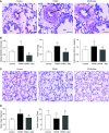
Figure 1.
Figure 1.
(A) Hematoxylin and eosin staining of lung sections showing pulmonary artery (PA) and adjacent airway (AW) in control, persistent pulmonary hypertension of the newborn (PPHN), and metformin-treated PPHN (PPHN + Met) lambs. Arrows point to PA lumen. Scale bars: 50 μm. (B) Summarized data for n = 4 lambs each for medial width/external diameter of pulmonary arteries in control, PPHN, and PPHN + Met lambs. Summarized data also shown for peak right ventricular systolic pressure (RVSP) and Fulton index for control, PPHN, and PPHN + Met lambs with n = 6 each for control, PPHN, and PPHN + Met. (C) Hematoxylin and eosin staining of lung sections showing alveolar structure in control, PPHN, and PPHN + Met groups. Scale bars: 50 μm. (D) PPHN lamb lung sections show alveolar simplification with a decrease in radial alveolar count (RAC) and increase in mean linear intercept (MLI). Summarized data are for n = 4 for each group. *Indicates P < 0.05 from control and #from PPHN groups by ANOVA and Tukey’s post hoc test. Metformin attenuated the increase in medial width, RVSP, and Fulton index in PPHN lambs. Metformin also restored alveolar growth with increase in RAC and decrease in MLI in PPHN lambs. LV = left ventricle; RV = right ventricle.
Figure 2.
Figure 2.
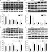
(A) Protein levels of AMPK, phosphorylated AMPK (p-AMPK), phosphorylated acetyl CoA carboxylase (p-ACC), its downstream target ACC, and mitochondrial transcription factor PGC-1α in control and PPHN cells by IB. Summarized data below are for samples from four lambs each, normalized to the protein levels of β-actin. (B) Protein levels of mitochondrial electron transport chain (ETC) complexes I–V, determined by IB. Summarized data below are for samples from four lambs each, normalized to protein levels of β-actin. (C and D) Effect of dominant negative AMPK construct (DN AMPK) on the levels of AMPK, p-AMPK, ACC, p-ACC, and PGC-1α (C) and mitochondrial ETC complex proteins I–V (D) in control pulmonary artery endothelial cells (PAEC). Normal PAEC were transfected with empty adeno virus (control) or adeno-DN AMPK. Increase in AMPK-α1 and presence of Myc tag verify the expression of DN AMPK in the transfected cells. Summarized data are for PAEC samples from three lambs normalized to β-actin. *Indicates P < 0.05 from controls by t test. Levels of p-AMPK, p-ACC, and PGC-1α, and mitochondrial ETC complexes were decreased in PPHN samples, whereas the levels of AMPK and ACC were not different. DN AMPK decreased the levels of p-AMPK, p-ACC, PGC-1α, and ETC complex proteins in control PAEC, reproducing the effects of PPHN. AMPK = 5′ AMP activated protein kinase.
Figure 3.
Figure 3.
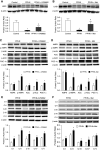
(A and B) Protein levels of LKB1 in control and PPHN PAEC and the effect of allosteric AMPK agonist A769662 (A) and metformin (B) on the LKB1 levels in PPHN PAEC. *Indicates P < 0.05 for LKB1 protein levels from control PAEC; #indicates P < 0.05 from saline-treated PPHN samples by ANOVA and Tukey’s post hoc test for n = 3 each. LKB1 levels were decreased in PPHN PAEC. Both A769662 and metformin increased the protein levels of LKB1 in PPHN PAEC. (C and D) The effects of A769662 (C) and metformin (D) on the levels of AMPK, p-AMPK, ACC, p-ACC, and PGC-1α. (E and F) Effects of A769662 (E) and metformin (F) on the mitochondrial ETC complex proteins I–V in PAEC from PPHN lambs. Summarized data are for samples from three control or three PPHN lambs treated with A769662 or metformin, normalized to protein levels of β-actin. *Indicates P < 0.05 from saline-treated PPHN samples for n = 3 by t test. A769662 and metformin increased the levels of p-AMPK, p-ACC, and PGC-1α and did not alter AMPK and ACC levels in PPHN PAEC. A769662 increased the levels of ETC complexes I–V and metformin increased the complexes I, II, III, and IV, in PPHN PAEC.
Figure 4.
Figure 4.
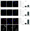
(A and B) Immunofluorescence staining for p-AMPK (A) and PGC-1α (B) in representative lung sections from control, PPHN, and metformin-treated PPHN lambs. Frozen lung sections from optimal cutting temperature compound inflated lungs were stained for p-AMPK and PGC-1α and for endothelial cell marker VE-cadherin to assess colocalization of these proteins with capillaries. Scale bars: 20 µm. Summarized data are for samples from four lambs in each group, showing integrated fluorescence density. *Indicates P < 0.05 from control lambs and #from PPHN lambs by ANOVA and Tukey’s post hoc test. Levels of p-AMPK and PGC-1α that colocalize to the blood vessels in the lung are decreased in PPHN. Metformin infusion in utero increased the fluorescence for p-AMPK and PGC-1α in the PPHN lamb lungs. (C) Representative confocal images of control, PPHN, and PPHN PAEC treated with 10−4 M metformin for 36 hours, with immunofluorescent labeling for TOM20 (translocase of outer mitochondrial membrane protein 20) and DAPI to outline nuclei with the corresponding black and white images shown below. Scale bars: 20 µm. Summarized data are from a total of three experiments. *Indicates P < 0.05 from control lambs and #from PPHN lambs by ANOVA and Tukey’s post hoc test. Mitochondrial number and networking were decreased in PPHN and were improved by metformin treatment of PPHN PAEC.
Figure 5.
Figure 5.
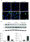
(A) Immunofluorescence staining and (B) IB for DLL4 and Jag1 in samples from control, PPHN and metformin-treated PPHN PAEC. Scale bars: 20 µm. Summarized data are for immunoblots done on samples from three control and three PPHN lambs each; PPHN + Met are PPHN cells treated with 10−4 M metformin for 48 hours. *Indicates P < 0.05 from control and #from PPHN-only samples. Expression of DLL4 was increased and Jag1 decreased in PPHN; metformin restored the expression of these notch ligands to control levels.
Figure 6.
Figure 6.
(A and B) In vitro tube formation assay in Matrigel for control and PPHN PAEC (A) and summarized data for measures of tube formation quantified by Image J angiogenesis analyzer (B). PAEC tube formation was assessed 4 to 6 hours after plating cells in Matrigel with Calcein staining. Summarized data are for control and PPHN cells obtained from three lambs each, treated with DN AMPK, A769662, metformin, Jag1 peptide, scrambled sequence Jag1 peptide (scrJag1), or DLL4 monoclonal antibody (DLL4 Mab). Scale bars: 800 μm. *Indicates P < 0.05 from control, #indicates P < 0.05 from PPHN alone for metformin, and from PPHN for A769669. For Jag1 and DLL4 Mab treatments, indicates P < 0.05 from PPHN + scrJag1 for Jag1 and from PPHN alone for DLL4 Mab treatment. DN AMPK decreased tube formation by control cells; metformin and A769662 did not alter tube formation by control cells. Tube formation was significantly impaired in PPHN PAEC. Both metformin and A769662 improved tube formation by PPHN PAEC. Jag1 peptide but not scrJag1, and DLL4 Mab have also improved tube formation by PPHN cells.
Figure 7.
Figure 7.
(A) Immunofluorescence staining for endothelial cell marker CD31 in frozen lung sections of optimal cutting temperature compound inflated lungs. Light DAPI (blue) staining was used to outline alveolar structure. Scale bars: 50 μm. Zoomed-in images of 20× magnification were shown in the second panel to show more detail for CD31 staining. Summarized data on the right shown for five lambs in each group. CD31-positive capillary number is decreased in PPHN lungs and was improved by metformin treatment of PPHN lambs in utero. (B) Pulmonary artery branching identified by micro–computed tomography angiography after Microfil perfusion of pulmonary artery. Summarized data to the right show mean vessel volume as a fraction of total lung volume for lung samples from five lambs in each group. Pulmonary artery branching was pruned, and vascular volume fraction was lower in PPHN lungs. Both were improved by metformin treatment in utero in PPHN lungs. *Indicates P < 0.05 from control and #from PPHN.

References

    1. Levin DL, Heymann MA, Kitterman JA, Gregory GA, Phibbs RH, Rudolph AM. Persistent pulmonary hypertension of the newborn infant. J Pediatr. 1976;89:626–630. - PubMed
    1. Haworth SG, Reid L. Persistent fetal circulation: newly recognized structural features. J Pediatr. 1976;88:614–620. - PubMed
    1. Grover TR, Parker TA, Balasubramaniam V, Markham NE, Abman SH. Pulmonary hypertension impairs alveolarization and reduces lung growth in the ovine fetus. Am J Physiol Lung Cell Mol Physiol. 2005;288:L648–L654. - PubMed
    1. Gien J, Seedorf GJ, Balasubramaniam V, Markham N, Abman SH. Intrauterine pulmonary hypertension impairs angiogenesis in vitro: role of vascular endothelial growth factor nitric oxide signaling. Am J Respir Crit Care Med. 2007;176:1146–1153. - PMC - PubMed
    1. Gien J, Seedorf GJ, Balasubramaniam V, Tseng N, Markham N, Abman SH. Chronic intrauterine pulmonary hypertension increases endothelial cell rho kinase activity and impairs angiogenesis in vitro. Am J Physiol Lung Cell Mol Physiol. 2008;295:L680–L687. - PMC - PubMed

Publication types

MeSH terms