. 2020 Feb 11;30(6):1670-1681.e7.
doi: 10.1016/j.celrep.2020.01.058.

NAD+ Repletion Rescues Female Fertility during Reproductive Aging

Affiliations

NAD+ Repletion Rescues Female Fertility during Reproductive Aging

Michael J Bertoldo et al. Cell Rep. .

Abstract

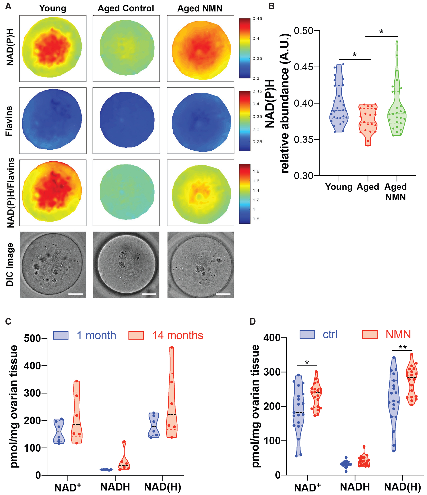
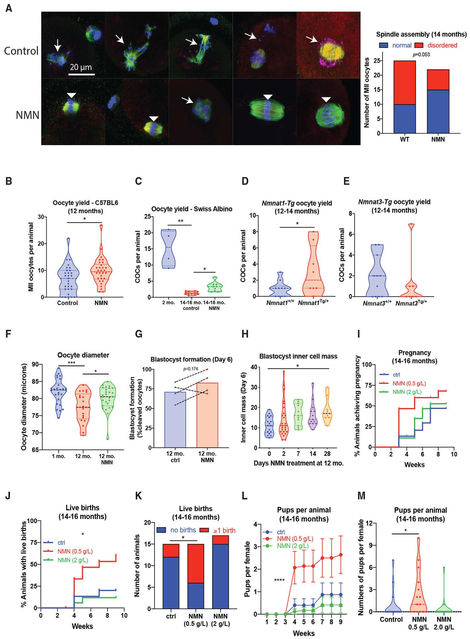
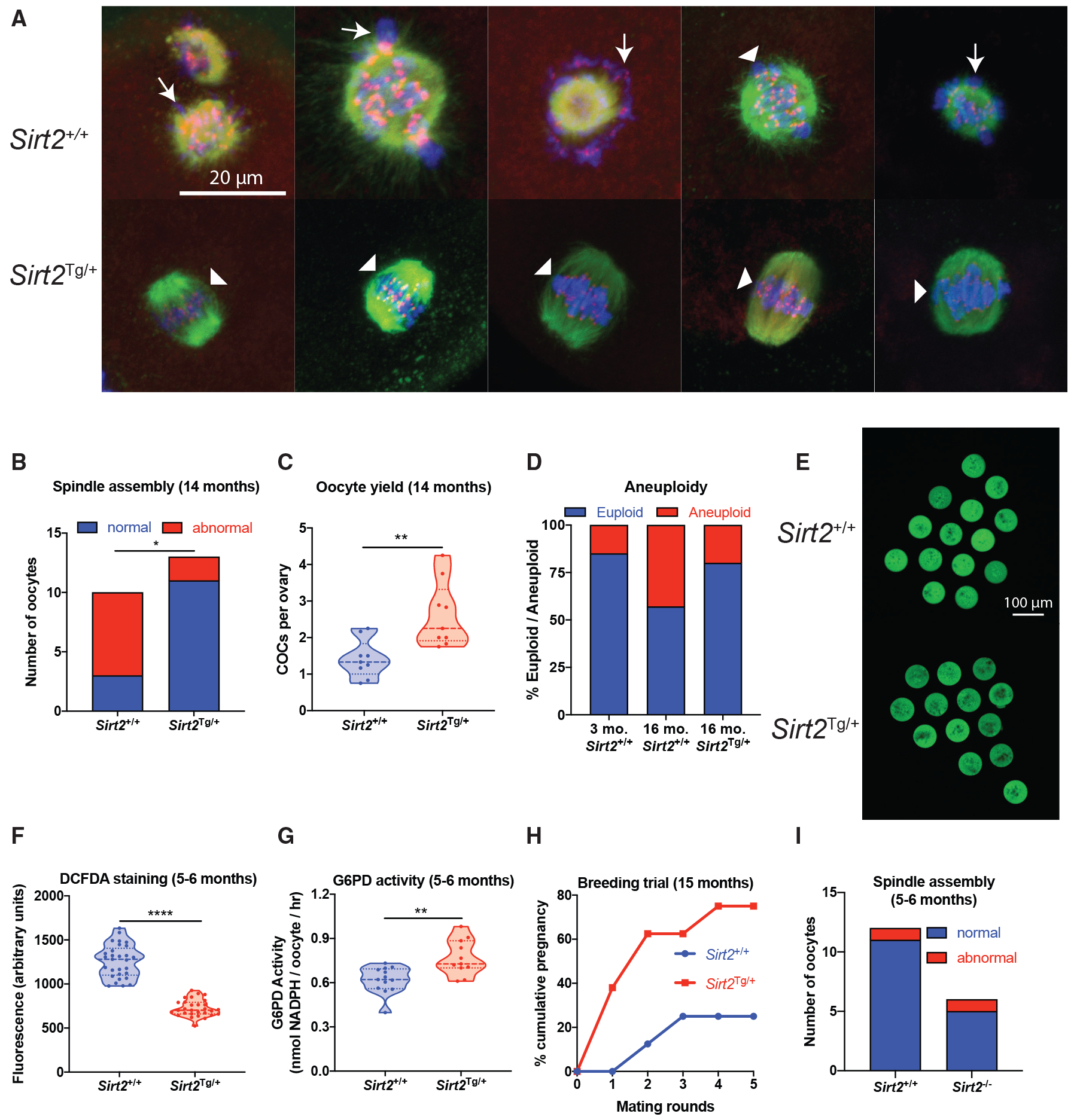
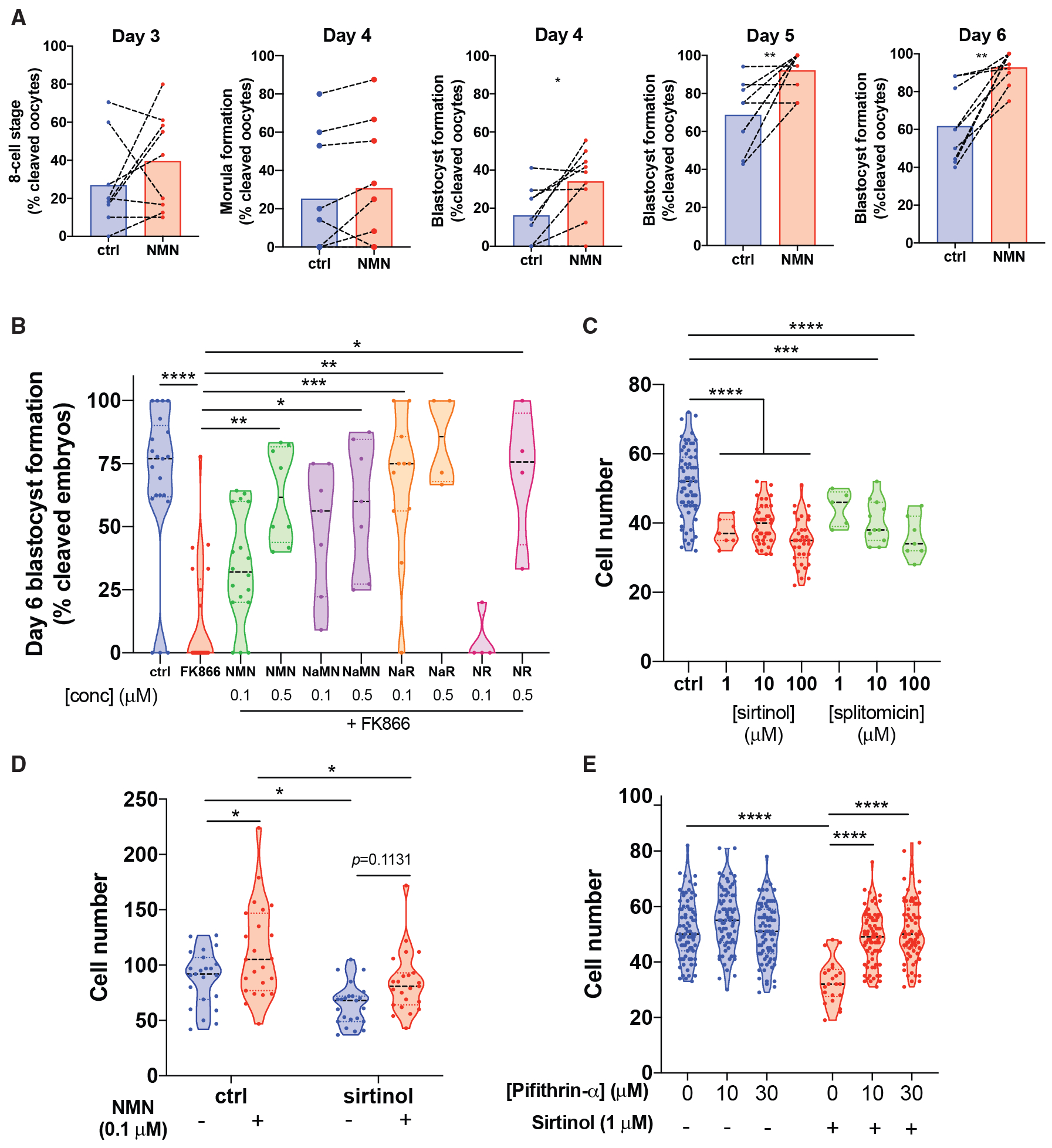
Reproductive aging in female mammals is an irreversible process associated with declining oocyte quality, which is the rate-limiting factor to fertility. Here, we show that this loss of oocyte quality with age accompanies declining levels of the prominent metabolic cofactor nicotinamide adenine dinucleotide (NAD+). Treatment with the NAD+ metabolic precursor nicotinamide mononucleotide (NMN) rejuvenates oocyte quality in aged animals, leading to restoration in fertility, and this can be recapitulated by transgenic overexpression of the NAD+-dependent deacylase SIRT2, though deletion of this enzyme does not impair oocyte quality. These benefits of NMN extend to the developing embryo, where supplementation reverses the adverse effect of maternal age on developmental milestones. These findings suggest that late-life restoration of NAD+ levels represents an opportunity to rescue female reproductive function in mammals.

Keywords: SIRT2; aging; embryo development; female fertility; in vitro fertilization; infertility; nicotinamide adenine dinucleotide (NAD+); nicotinamide mononucleotide (NMN); oocyte; reproductive aging.


Conflict of interest statement

Declaration of Interests L.E.W., H.A.H., and D.A.S. are cofounders, shareholders, directors, and advisers of Jumpstart Fertility Inc., which was founded to develop the work described here, and are inventors on a patent (WO2019023748A1 “Methods to improve fertility”) that forms the basis of this work and has been licensed to Jumpstart Fertility. D.A.S. is an inventor on a patent (WO2013002880A1) that was previously licensed to Jumpstart Fertility. The salaries of M.J.B. and D.M.G. working in the labs of L.E.W. and R.B.G. at UNSW were paid by sponsored research from Jumpstart Fertility to UNSW. K.S. was an employee of Jumpstart Fertility. W.-H.J.H. and D.R.L. hold shares in Jumpstart Fertility. H.A.H. undertakes ART in the private sector in affiliation with Queensland Fertility Group. L.E.W. and D.A.S. are also advisers and shareholders in EdenRoc Sciences (Metro Biotech NSW, Metro Biotech, Liberty Biosecurity) and in Life Biosciences LLC and its daughter companies (Jumpstart Fertility, Continuum Biosciences, Senolytic Therapeutics, Selphagy, Animal Biosciences, Iduna). L.E.W. provides consulting work for Life Biosciences and is an adviser and shareholder in Intravital Pty Ltd. D.A.S. is also a founder, equity owner, adviser, director, consultant, investor, and/or inventor on patents licensed to Vium, Jupiter Orphan Therapeutics, Cohbar, Galilei Biosciences, Wellomics, EdenRoc Sciences (and affiliates Arc-Bio, Dovetail Genomics, Claret, Revere Biosciences, UpRNA, MetroBiotech, Liberty Biosecurity), Life Biosciences (and affiliates Selphagy, Senolytic Therapeutics, Spotlight Therapeutics, Animal Biosciences, Iduna, Continuum, Jumpstart Fertility, Iduna). D.A.S. is an inventor on a patent application filed by Mayo Clinic and Harvard Medical School that has been licensed to Elysium Health. For details see https://genetics.med.harvard.edu/sinclair.

Figures

Figure 1.
Figure 1.. Oocyte NAD+ Levels Decline with Age and Are Increased with NMN Treatment
(A and B) Multispectral imaging (A) to determine NAD(P)H content in oocytes from young (4- to 5-week-old) or aged (12-month-old) mice treated with NMN (drinking water, 2 g/L, 4 weeks) (scale bar is 20 μm), quantified in (B) (one-way ANOVA 1.454 (2, 71), *p = 0.0165, 0.0287 by Dunnett’s multiple comparison test, n = 21–27 oocytes per group). (C and D) Mass spectrometry of the whole ovary (C) shows no change in NAD(H) levels between the ages of 1 and 14 months (n = 6 mice per group); however, (D) NMN increased ovarian NAD(H) in 14-month-old mice (1 h following 400 mg/kg oral gavage, *p = 0.0123, **p = 0.0072, two-tailed t test, n = 20 mice per group).
Figure 2.
Figure 2.. NMN Treatment Restores Oocyte Quality, Follicle Dynamics, Embryo Development, and Live Birth Rates
(A) NMN treatment (drinking water, 2 g/L, 4 weeks) with NMN from 14 months restores spindle assembly in immunostained oocytes (β-tubulin in green, Hoechst for DNA in blue, kinetochores in red, p = 0.0503 by Fisher’s exact test, n = 23–25 per group). Disordered spindles with lagging chromosomes are indicated by arrows, and normal, barrel-shaped bipolar spindles with DNA aligned along the metaphase plate are indicated by arrowheads. (B and C) Oocyte yield following ovarian stimulation in (B) aged (12-month-old) C57BL/6JAusb mice (*p = 0.0211, two-tailed t-test, n = 29–30 animals per group) and (C) 14- to 16-month-old Swiss albino mice (Kruskal-Wallis 16.31, ***p = 0.0004, *p = 0.0295 by Dunnett’s mulitple comparison test, n = 4–11 per group). (D and E) Aged (12- to 14-month-old) transgenic mice overexpressing NMNAT1 have increased oocyte yield (*p = 0.0416, two-tailed t-test, n = 8–10 per group) (D) in comparison to transgenics overexpressing NMNAT3 (n = 7–11) (E). (F) Oocyte diameter following NMN treatment (2 g/L, drinking water, 4 weeks) in aged (12-month-old) or young (4- to 6-week-old) C57BL/6 females (F(2,71) = 8.504, ***p = 0.0002, *p = 0.0332 by Dunnett’s multiple comparison test, n = 21–27 oocytes per group). (G) In a parallel cohort, oocytes were used for IVF, with blastocyst formation at day 6 of embryo development (each datapoint is cumulative data from one independent experiment). (H) 12-month-old C57BL/6 females were treated for the indicated times with NMN in drinking water (2 g/L), and MII oocytes were subjected to IVF. At day 6, inner cell mass was assessed (Kruskal-Wallis 11.93, *p = 0.0337 by Dunn’s multiple comparison test, n = 8–26 oocytes per group). Previously untreated 13-month-old C57BL/6 females (n = 15–17 per group) were treated for 4 weeks with NMN in drinking water at two doses (0.5 and 2 g/L), following which a male was introduced and breeding performance was assessed over the next 9 weeks. (I–M) Breeding performance was assessed by (I) cumulative time to pregnancy, (J) cumulative time to live birth (log-rank test, **p = 0.0059), (K) overall proportion achieving live birth (Fisher’s exact test, *p = 0.0253 ctrl versus 0.5 g/L NMN), (L) cumulative number of pups born over time (repeated-measures ANOVA, NMN F(2, 43) = 4.925, p = 0.0119, Dunnett’s multiple comparison ctrl versus NMN 0.5, ****p < 0.0001, error bars show SEM), and (M) overall number of pups per female (Kruskal-Wallis 9.220, p = 0.0100, Dunn’s multiple comparison, *p = 0.0491 ctrl versus 0.5 g/L NMN).
Figure 3.
Figure 3.. SIRT2 Transgenic Mice Have Improved Oocyte Quality
(A and B) Oocytes from 14-month-old Sirt2Tg/+ C57BL/6 mice were subjected to (A) immunostaining for spindle assembly (β-tubulin in green, Hoechst for DNA in blue, kinetochores in red). Disordered spindles with lagging chromosomes are indicated by arrows, and normal, barrel-shaped bipolar spindles with DNA aligned along the metaphase plate are indicated by arrowheads; this is quantified in (B) (p = 0.0131, n = 10–13 oocytes per group, Fisher’s exact test). (C) Oocyte yield from aged (14-month-old) Sirt2Tg/+ and wild-type Sirt2+/+ littermates (**p = 0.0030, two-tailed t-test, n = 9 animals per group). (D) Aneuploidy rates in oocytes from young (2-month-old) and aged (16-month-old) Sirt2Tg/+ and Sirt2+/+ littermates (n = 26 young, n = 5–7 aged). (E–G) Oocytes from Sirt2Tg/+ mice had decreased ROS levels as determined by (E) H2DCFDA staining, quantified in (F) (****p < 0.0001, two-tailed t-test, n = 29 oocytes per group), with (G) increased G6PD enzyme activity (**p = 0.0019, two-tailed t-test, n = 11–13 using 5 pooled oocytes per sample from 4 animals per group). (H) Mating trials from 15 months of age to determine cumulative pregnancy rates (p = 0.1319 after 5 mating rounds, n = 8 animals per group, Fisher’s exact test). (I) Spindle assembly in oocytes from Sirt2−/− knockout animals (n = 6, 14 oocytes per group).
Figure 4.
Figure 4.. In Vitro NMN Treatment Enhances Embryo Formation
(A) Oocytes from aged (12-month-old) mice were subjected to IVF, and embryos maintained in 1 μM NMN until day 6 of embryo development (paired two-tailed t test, day 4 *p = 0.0138, day 5 **p = 0.0086, day 6 **p = 0.0014, n = 9 independent experiments). (B) Treatment with the NAMPT inhibitor FK866 causes embryo death at day 6, which can be rescued by the NAD+ precursors NMN, nicotinic acid mononucleotide (NaMN), nicotinic acid riboside (NaR), and NR (Kruskal-Wallis 50.65, p < 0.0001, ****p < 0.0001, **p = 0.0048, *p = 0.0197, ***p = 0.0004, **p = 0.0014, *p = 0.0156, n = 4–21 embryos per group as shown). (C) Treatment with sirtinol or splitomicin inhibits blastocyst formation (supplemental), with decreased cell count in blastocysts (one-way ANOVA F(6,162) = 22.44, ***p = 0.0002, ****p < 0.0001 by Dunnett’s multiple comparison test, n = 5–67 per group as shown by raw datapoints). (D) Co-treatment of sirtinol-treated embryos with NMN rescues this reduction (two-way ANOVA F(1,88) sirtinol = 17.66, NMN = 12.59, *p = 0.0360, 0.0374, ****p < 0.0001 by Sidak’s multiple comparison test, n = 22–23 per group). (E) Treatment with the p53 inhibitor pifithrin rescues cell count and blastocyst formation (supplemental) in embryos treated with sirtinol (pifithrin F(2,392) = 22.37,0020sirtinol F(1,392) = 49.81, ****p < 0.0001 by Sidak’s multiple comparison test, n = 72–78 per group).

References

    1. Agarwal A, Aponte-Mellado A, Premkumar BJ, Shaman A, and Gupta S (2012). The effects of oxidative stress on female reproduction: a review. Reprod. Biol. Endocrinol 10, 49. - PMC - PubMed
    1. Atzmon Y, Shoshan-Karchovsky E, Michaeli M, Aslih N, Shrem G, Ellenbogen A, and Shalom-Paz E (2017). Obesity results with smaller oocyte in in vitro fertilization/intracytoplasmic sperm injection cycles-a prospective study. J. Assist. Reprod. Genet 34, 1145–1151. - PMC - PubMed
    1. Bedalov A, Gatbonton T, Irvine WP, Gottschling DE, and Simon JA (2001). Identification of a small molecule inhibitor of Sir2p. Proc. Natl. Acad. Sci. USA 98, 15113–15118. - PMC - PubMed
    1. Berger F, Lau C, Dahlmann M, and Ziegler M (2005). Subcellular compartmentation and differential catalytic properties of the three human nicotinamide mononucleotide adenylyltransferase isoforms. J. Biol. Chem 280, 36334–36341. - PubMed
    1. Bieganowski P, and Brenner C (2004). Discoveries of nicotinamide riboside as a nutrient and conserved NRK genes establish a Preiss-Handler independent route to NAD+ in fungi and humans. Cell 117, 495–502. - PubMed

Publication types

LinkOut - more resources