

CBCT assessment of bone thickness in maxillary and mandibular teeth: an anatomic study
- PMID: 32049133
- PMCID: PMC6999116
- DOI: 10.1590/1678-7757-2019-0148
CBCT assessment of bone thickness in maxillary and mandibular teeth: an anatomic study
Abstract
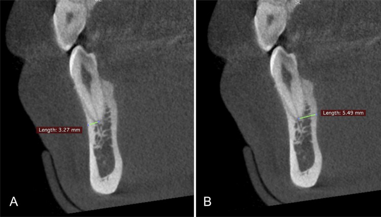
Objective: The site of the sinus tract depends on the rate of resistance against abscess exudate drainage, bone morphology, and distance from the root apex to the outer cortical bone. To assess apical bone thickness in buccal and palatal/lingual aspects of maxillary and mandibular teeth, using a high-resolution cone-beam computed tomography (CBCT) system.
Methodology: In total, 422 CBCT examinations were included in the study, resulting in a sample of 1400 teeth. The scans were acquired by PreXion 3D, with a high-resolution protocol. The bone thickness was taken as the distance between the center of the apical foramen and the buccal and lingual/palatal cortical bone. The quantitative variables were expressed as mean values±standard deviation. The independent samples were analyzed using the t-test or the Mann-Whitney test (p<0.05).
Results: The lowest mean value of bone thickness was observed in the buccal cortical bone of the upper canines (1.49 mm±0.86) and in the upper central incisors (1.59 mm±0.67). In premolar teeth, the lowest values were found in the buccal cortical bone of upper first premolars (1.13 mm±0.68). In the posterior teeth, the lowest values were found in the buccal cortical bone of upper first molars (1.98 mm±1.33). In the lower second molar region, the buccal cortical bone (8.36 mm±1.84) was thicker than the lingual cortical bone (2.95 mm±1.16) (p<0.05).
Conclusions: The lowest mean values of bone thickness are in the buccal cortical bone of the maxillary teeth. In the mandible, bone thickness is thinner in the buccal bone around the anterior and premolar teeth, and in the lingual aspect of mandibular molars. All these anatomic characteristics could make the occurrence of the sinus tract more susceptible in these specific regions of the maxillary and mandibular alveolar bone.
Figures


References
-
- - Schulz M, von Arx T, Altermatt HJ, Bosshardt D. Histology of periapical lesions obtained during apical surgery. J Endod. 2009;35(5):634-42. - PubMed
- Schulz M, von Arx T, Altermatt HJ, Bosshardt D. Histology of periapical lesions obtained during apical surgery. J Endod. 2009;35(5):634–642. - PubMed
-
- - Ricucci D, Loghin S, Gonçalves LS, Rôças IN, Siqueira JF Jr. Histobacteriologic conditions of the apical root canal system and periapical tissues in teeth associated with sinus tracts. J Endod. 2018;44(3):405-13. - PubMed
- Ricucci D, Loghin S, Gonçalves LS, Rôças IN, Siqueira JF., Jr Histobacteriologic conditions of the apical root canal system and periapical tissues in teeth associated with sinus tracts. J Endod. 2018;44(3):405–413. - PubMed
-
- - Miri SS, Atashbar O, Atashbar F. Prevalence of sinus tract in the patients visiting department of endodontics, Kermanshah School of Dentistry. Glob J Health Sci. 2015;7(6):271-5. - PMC - PubMed
- Miri SS, Atashbar O, Atashbar F. Prevalence of sinus tract in the patients visiting department of endodontics, Kermanshah School of Dentistry. Glob J Health Sci. 2015;7(6):271–275. - PMC - PubMed
-
- - López-Jarana P, Díaz-Castro CM, Falcão A, Falcão C, Ríos-Santos JV, Herrero-Climent M. Thickness of the buccal bone wall and root angulation in the maxilla and mandible: an approach to cone beam computed tomography. BMC Oral Health. 2018;18(1):194. - PMC - PubMed
- López-Jarana P, Díaz-Castro CM, Falcão A, Falcão C, Ríos-Santos JV, Herrero-Climent M. Thickness of the buccal bone wall and root angulation in the maxilla and mandible: an approach to cone beam computed tomography. 194BMC Oral Health. 2018;18(1) - PMC - PubMed
MeSH terms
LinkOut - more resources
Full Text Sources
Miscellaneous
