. 2020 Feb 7;61(2):14.
doi: 10.1167/iovs.61.2.14.

nGnG Amacrine Cells and Brn3b-negative M1 ipRGCs are Specifically Labeled in the ChAT-ChR2-EYFP Mouse

Affiliations

nGnG Amacrine Cells and Brn3b-negative M1 ipRGCs are Specifically Labeled in the ChAT-ChR2-EYFP Mouse

Ling-Jie Cui et al. Invest Ophthalmol Vis Sci. .

Abstract

Purpose: Experimental access to specific cell subtypes is essential for deciphering the complexity of retinal networks. Here, we characterized the selective labeling, caused by ectopic transgene expression, of two atypical retinal neurons in the ChAT-Channelrhodopsin-2 (ChR2)-EYFP mouse.

Methods: Retinal sections and flat-mounts were prepared for double-staining immunohistochemistry with antibodies against EYFP and various neuronal markers. Sagittal/coronal brain slices were made to visualize EYFP signals in central nuclei. Whole-cell recordings were conducted to test the functionality of ChR2.

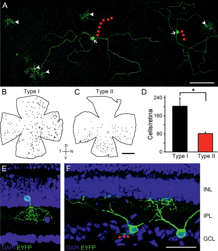
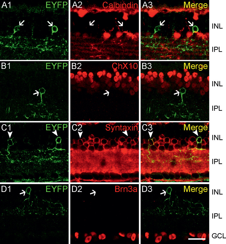
Results: Two populations of EYFP-positive retinal cells were observed. The inner nuclear layer (INL)-located one (type I cell) distributed regularly throughout the entire retina, whereas the ganglion cell layer (GCL)-residing one (type II cell) was restricted ventrally. None of them was cholinergic, as evidenced by the complete absence of ChAT immunoreactivity. Type I cells were immunolabeled by the amacrine marker syntaxin. However, the vast majority of them were neither positive to GABA/GAD65, nor to GlyT1/glycine, suggesting that they were non-GABAergic non-glycinergic amacrine cells (nGnG ACs), which was confirmed by double-labeling with the nGnG AC marker PPP1R17. Type II cells were immunopositive to melanopsin, but not to Brn3a or Brn3b. They possessed dendrites stratifying in the outermost inner plexiform layer (IPL) and axons projecting to the suprachiasmatic nucleus (SCN) rather than the olivary pretectal nucleus (OPN), suggesting that they belonged to a Brn3b-negative subset of M1-type intrinsically photosensitive retinal ganglion cells (ipRGCs). Glutamatergic transmission-independent photocurrents were elicited in EYFP-positive cells, indicating the functional expression of ChR2.

Conclusions: The ChAT-ChR2-EYFP retina exhibits ectopic, but functional, transgene expression in nGnG ACs and SCN-innervating M1 ipRGCs, thus providing an ideal tool to achieve efficient labeling and optogenetic manipulation of these cells.


Conflict of interest statement

Disclosure: L.-J. Cui, None; W.-H. Chen, None; A.-L. Liu, None; X. Han, None; S.-X. Jiang, None; F. Yuan, None; Y.-M. Zhong, None; X.-L. Yang, None; S.-J. Weng, None

Figures

Figure 1.
Figure 1.
General expression profile of the transgene in the ChAT-ChR2-EYFP retina. (A) Micrograph of a whole-mount ChAT-ChR2-EYFP retina labeled with an antibody directed to EYFP, which reveals EYFP expression in two cell populations with distinct morphologies: type I cells (white arrowheads) with small somata and narrow, heavily branched dendritic fields and type II cells (white arrows) with large cell bodies and wide-field, sparely branched dendrites. Red arrows point to vaguely labeled axons. Scale bar = 90 µm. (B, C) Representative silhouettes showing topographic distributions of type I (B) and II (C) cells. Scale bar = 1 mm. D, dorsal; V, ventral; T, temporal; N, nasal. (D) Bar chart summarizing the average number of types I and II cells per retina. Error bars represent 1 SEM. (E, F) Representative confocal fluorescence microphotographs of retinal vertical sections, showing the localization of somata and dendrites of a type I (E) and two type II (F) cells. Nuclei were counterstained with DAPI (blue). The type I cell has a soma located in the proximal INL and processes arborizing diffusely across the sublaminae S1-S3 of the IPL. The type II cells have somata located in the GCL and processes ramifying at the outermost IPL. An axon can be seen (red arrows). Scale bar = 40 µm.
Figure 2.
Figure 2.
Lack of ChAT immunoreactivity in EYFP-positive retinal cells. (A1B3) Representative micrographs captured from whole-mount retinas double-labeled for EYFP (left panels) and ChAT (middle panels). ChAT labeling is not seen in EYFP-positive neurons in either the INL (A1A3, type I cells) or GCL (B1B3, type II cells). Arrows indicate EYFP-labeled, but ChAT-negative cells. Scale bar = 20 µm.
Figure 3.
Figure 3.
EYFP immunoreactivity in the INL is localized to amacrine cells. EYFP-expressing retinal sections were immunostained for the horizontal cell marker calbindin (A1A3), bipolar cell marker Chx10 (B1B3), amacrine cell marker syntaxin (C1C3) and ganglion cell marker Brn3a (D1D3). As observed in the merged images (right panels), EYFP-positive cells lack any detectable immunoreactivity for calbindin, Chx10, or Brn3a (arrows), but are immunopositive to syntaxin (arrowheads). Scale bar = 20 µm.
Figure 4.
Figure 4.
EYFP immunoreactivity in the INL is localized to nGnG amacrine cells but not to GABAergic or glycinergic amacrine cells. Retinal sections were co-stained with EYFP and biomarkers for three major classes of ACs, including: GABAergic makers GABA (A1A3) and GAD65 (B1B3), glycinergic markers GlyT1 (C1C3), and glycine (D1D3), and PPP1R17, a specific marker for nGnG ACs (E1E3). From the merged images (right panels), it is clear that virtually no immunosignals for GABAergic or glycinergic markers are seen in EYFP-positive cells (arrows) in the INL. By contrast, all EYFP-positive cells exhibit robust PPP1R17 immunoreactivity (arrowheads). Scale bar = 20 µm.
Figure 5.
Figure 5.
Absence of immunoreactivity for various conventional amacrine markers in EYFP-positive INL cells. Vertical sections prepared from ChAT-ChR2-EYFP retinas were co-immunolabeled with antibodies probing EYFP and TH (dopaminergic AC marker, A1A3), VIP (VIP-expressing AC marker, B1B3), bNOS (bNOS-expressing AC marker, C1C3), VGluT3 (glutamatergic AC marker, D1D3), and VGluT1 (glutamatergic neuronal marker, E1E3). Note that virtually no EYFP-positive INL cells are co-labeled by any of the markers listed above. In all the panels, arrows point to EYFP-positive cells, and all these cells were negative to markers listed. TH, tyrosine hydroxylase; VIP, vasoactive intestinal polypeptide; bNOS, nitric oxide synthase, brain (251–270); VGluT3, vesicular glutamate transporter 3; VGluT1, vesicular glutamate transporter 1. Scale bars = 40 µm in A3 (applies to A1A3) and 20 µm in E3 (applies to B1E3).
Figure 6.
Figure 6.
EYFP immunoreactivity in the GCL is localized to M1 ipRGCs. (A1C3) Representative micrographs of retinal whole-mounts showing that EYFP-positive somata residing in the GCL lack immunoreactivity of Brn3a (GC marker, arrows in A1A3), but are stained by two melanopsin antibodies, UF006 (marker of various ipRGC subtypes, arrowheads in B1B3) and PA1-780 (M1-type ipRGC marker, arrowheads in C1C3). Scale bar = 20 µm. (D) Examples demonstrating the morphological profiles of EYFP-positive GCs as reconstructed from whole-mount retinas. The large, sparsely branched dendritic field is similar among these cells, typical of M1-type ipRGCs. Arrowheads indicate axons. Scale bar = 60 µm.
Figure 7.
Figure 7.
Axonal terminations of EYFP-positive M1 ipRGCs in the SCN of the hypothalamus. (A1A3) Representative micrographs of a retinal whole-mount double-stained for EYFP (left) and Brn3b (middle), showing that a GCL-residing EYFP-positive cell (arrow) is Brn3b-negative (right). Scale bar = 20 µm. (B1C3) Coronal brain sections in which retinal projections were labeled by intraocular injection of Alexa Fluor 594-conjugated CTB. Outlines of the SCN and OPN, two NIF visual centers known to receive extensive innervations from M1 ipRGCs, are depicted with white dots. EYFP-positive fibers are enriched in the SCN (B1B3), whereas very few, if any, EYFP signals are observed in the OPN (C1C3). Scale bar = 125 µm.
Figure 8.
Figure 8.
Functional ChR2 is expressed by EYFP-positive cells. (A1B3) Confocal photomicrographs of retinal sections co-stained with antibodies recognizing EYFP and ChR2. Robust ChR2 signals are co-localized to EYFP fluorescence in both type I (A1A3) and type II cells (B1B3). Arrows point to somata. Scale bar = 20 µm. (C1) Representative infrared image of a whole-mount retina, in which a type I cell (white arrow) was recorded with a micropipette (red arrowheads). (C2) A fluorescent image taken in the same area in C1, indicating the EYFP-positive soma of the cell recorded. Scale bar = 20 mm. (C3) Voltage-clamp recordings of an inward photocurrent (Vhold: 67 mV), elicited from the cell shown in C1 and C2, in response to full-field light stimulation in the presence of a glutamatergic blocker cocktail (L-AP4, DNQX, ACET and D-AP5). The blue bar represents the 470 nm light pulse.

References

    1. Masland RH. Neuronal diversity in the retina. Curr Opin Neurobiol. 2001; 11: 431–436. - PubMed
    1. Masland RH. The neuronal organization of the retina. Neuron. 2012; 76: 266–280. - PMC - PubMed
    1. Baden T, Berens P, Franke K, Roman Roson M, Bethge M, Euler T. The functional diversity of retinal ganglion cells in the mouse. Nature. 2016; 529: 345–350. - PMC - PubMed
    1. Bae JA, Mu S, Kim JS, et al. .. Digital museum of retinal ganglion cells with dense anatomy and physiology. Cell. 2018; 173: 1293–1306e1219. - PMC - PubMed
    1. Rheaume BA, Jereen A, Bolisetty M, et al. .. Single cell transcriptome profiling of retinal ganglion cells identifies cellular subtypes. Nat Commun. 2018; 9: 2759. - PMC - PubMed

Publication types