. 2020 May 15;26(28):6214-6223.
doi: 10.1002/chem.201905272. Epub 2020 Apr 28.

Mechanistic Insights into the Regio- and Stereoselectivities of Testosterone and Dihydrotestosterone Hydroxylation Catalyzed by CYP3A4 and CYP19A1

Affiliations

Mechanistic Insights into the Regio- and Stereoselectivities of Testosterone and Dihydrotestosterone Hydroxylation Catalyzed by CYP3A4 and CYP19A1

Junhao Li et al. Chemistry. .

Abstract

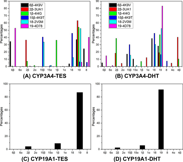
The hydroxylation of nonreactive C-H bonds can be easily catalyzed by a variety of metalloenzymes, especially cytochrome P450s (P450s). The mechanism of P450 mediated hydroxylation has been intensively studied, both experimentally and theoretically. However, understanding the regio- and stereoselectivities of substrates hydroxylated by P450s remains a great challenge. Herein, we use a multi-scale modeling approach to investigate the selectivity of testosterone (TES) and dihydrotestosterone (DHT) hydroxylation catalyzed by two important P450s, CYP3A4 and CYP19A1. For CYP3A4, two distinct binding modes for TES/DHT were predicted by dockings and molecular dynamics simulations, in which the experimentally identified sites of metabolism of TES/DHT can access to the catalytic center. The regio- and stereoselectivities of TES/DHT hydroxylation were further evaluated by quantum mechanical and ONIOM calculations. For CYP19A1, we found that sites 1β, 2β and 19 can access the catalytic center, with the intrinsic reactivity 2β>1β>19. However, our ONIOM calculations indicate that the hydroxylation is favored at site 19 for both TES and DHT, which is consistent with the experiments and reflects the importance of the catalytic environment in determining the selectivity. Our study unravels the mechanism underlying the selectivity of TES/DHT hydroxylation mediated by CYP3A4 and CYP19A1 and is helpful for understanding the selectivity of other substrates that are hydroxylated by P450s.

Keywords: C−H activation; P450; density functional calculations; hydroxylation; molecular modeling; steroids.


Conflict of interest statement

The authors declare no conflict of interest.

Figures

Figure 1
Figure 1
Chemical structures of TES and DHT and the corresponding SOMs. The bonds connecting the α hydrogen atoms are represented as dashed lines. The SOMs of TES/DHT by CYP3A4 and CYP19A1 are colored in red and blue, respectively; the SOM that can be hydroxylated by both CYP3A4 and CYP19A1 is colored in cyan.
Figure 2
Figure 2
Binding modes predicted by molecular docking. A) The three representative 17‐OHUP binding modes: 194D78 (magenta), 1β4I4G (yellow) and 2β3UA1 (green); B) The three representative 17‐OHDOWN binding modes: 6β4K9V (yellow), 15β4Κ9Τ (green) and 182V0M (magenta).
Figure 3
Figure 3
Accessibility profiles from the MD simulations. The percentage for a site is the number of snapshots in which the site was accessed divided by the number of snapshots for all the accessed sites.
Figure 4
Figure 4
A) TS structures for sites 19 (in cyan) and 6β (in magenta) of TES in the 194D78 system. B) The TS structures for sites 19 (in cyan) and 4β (in green) of DHT in the 194D78 system. C) The TS structures for sites 19 (in cyan), 6β (in magenta) and 8 (in blue) of TES in the 6β4K9V system. D) The TS structures for sites 19 (in cyan), 18 (in yellow), and 8 (in blue) of DHT in the 6β4K9V system. E) The TS structures for sites 19 (in cyan), 1β (in light pink) and 2β (in blue) of TES in the CYP19A1 system. F) The TS structures for sites 19 (in cyan), 1β (in light pink) and 2β (in blue) of DHT in the CYP19A1 system. The activation barrier (in kcal mol−1) for each site is shown in parentheses. The Habstracted−oxo and Habstracted−Csite distances are in Å.
Figure 5
Figure 5
Spin natural orbital (SNO) distributions for the TS structures for site 19 in the CYP3A4‐DHT systems: A) the α electron density in 17‐OHUP; B) the β electron density in 17‐OHUP; C) the α electron density in 17‐OHDOWN; D) the β electron density in 17‐OHDOWN. The SNOs were generated by the Multiwfn 3.6 package30 and visualized using VMD 1.9.331 with isovalue=0.02.
Figure 6
Figure 6
Spin natural orbital (SNO) distributions for the TS structures for site 19 in the CYP19A1 systems: A) the α electron density in CYP19A1‐TES; B) the β electron density in CYP19A1‐TES; C) the α electron density in CYP19A1‐DHT; D) the β electron density in CYP19A1‐DHT. The SNOs were generated by the Multiwfn 3.6 package30 and visualized using VMD 1.9.331 with isovalue=0.02.

References

    1. None
    1. Denisov I. G., Makris T. M., Sligar S. G., Schlichting I., Chem. Rev. 2005, 105, 2253–2278; - PubMed
    1. Guengerich F. P., Chem. Res. Toxicol. 2008, 21, 70–83. - PubMed
    1. None
    1. Meunier B., de Visser S. P., Shaik S., Chem. Rev. 2004, 104, 3947–3980; - PubMed

LinkOut - more resources