. 2020 Feb 12;14(2):e0008027.
doi: 10.1371/journal.pntd.0008027. eCollection 2020 Feb.

A high infectious simian adenovirus type 23 vector based vaccine efficiently protects common marmosets against Zika virus infection

Affiliations

A high infectious simian adenovirus type 23 vector based vaccine efficiently protects common marmosets against Zika virus infection

Shengxue Luo et al. PLoS Negl Trop Dis. .

Abstract

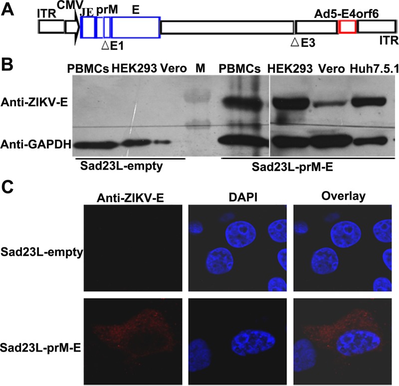
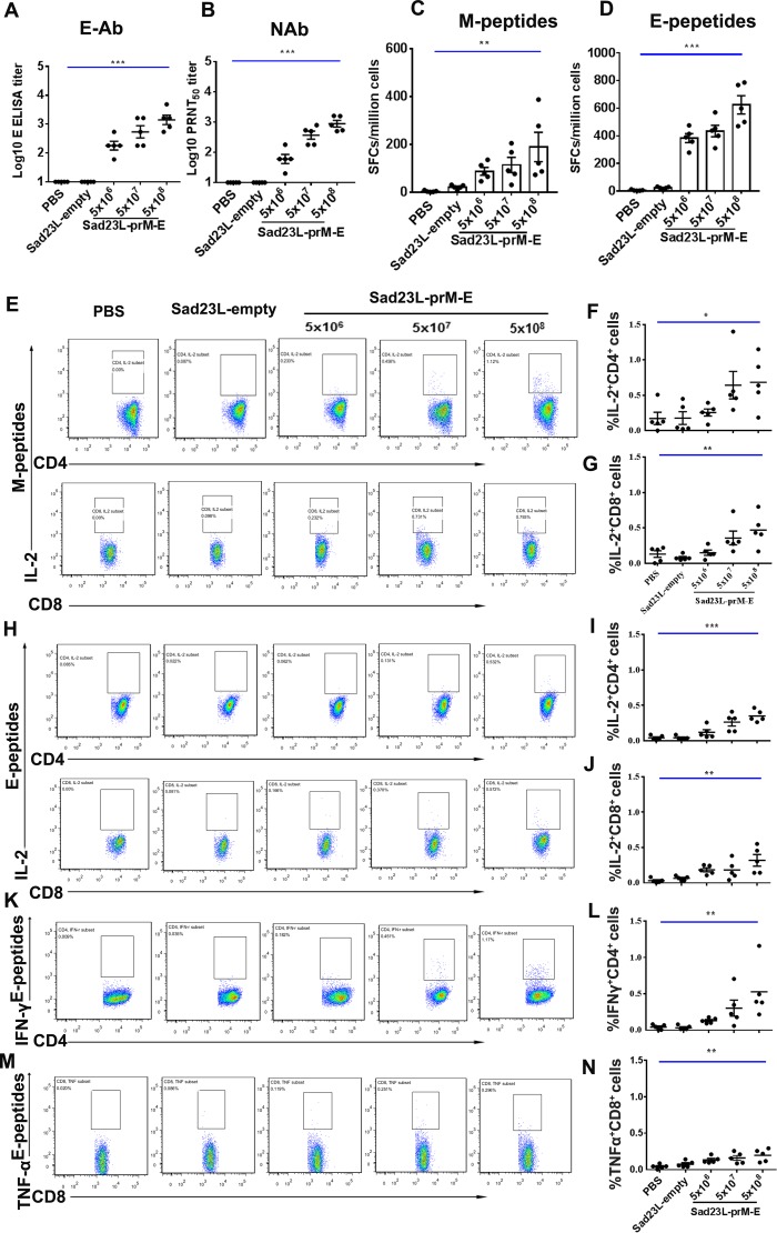
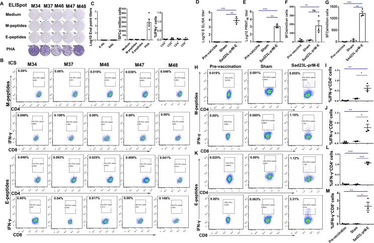
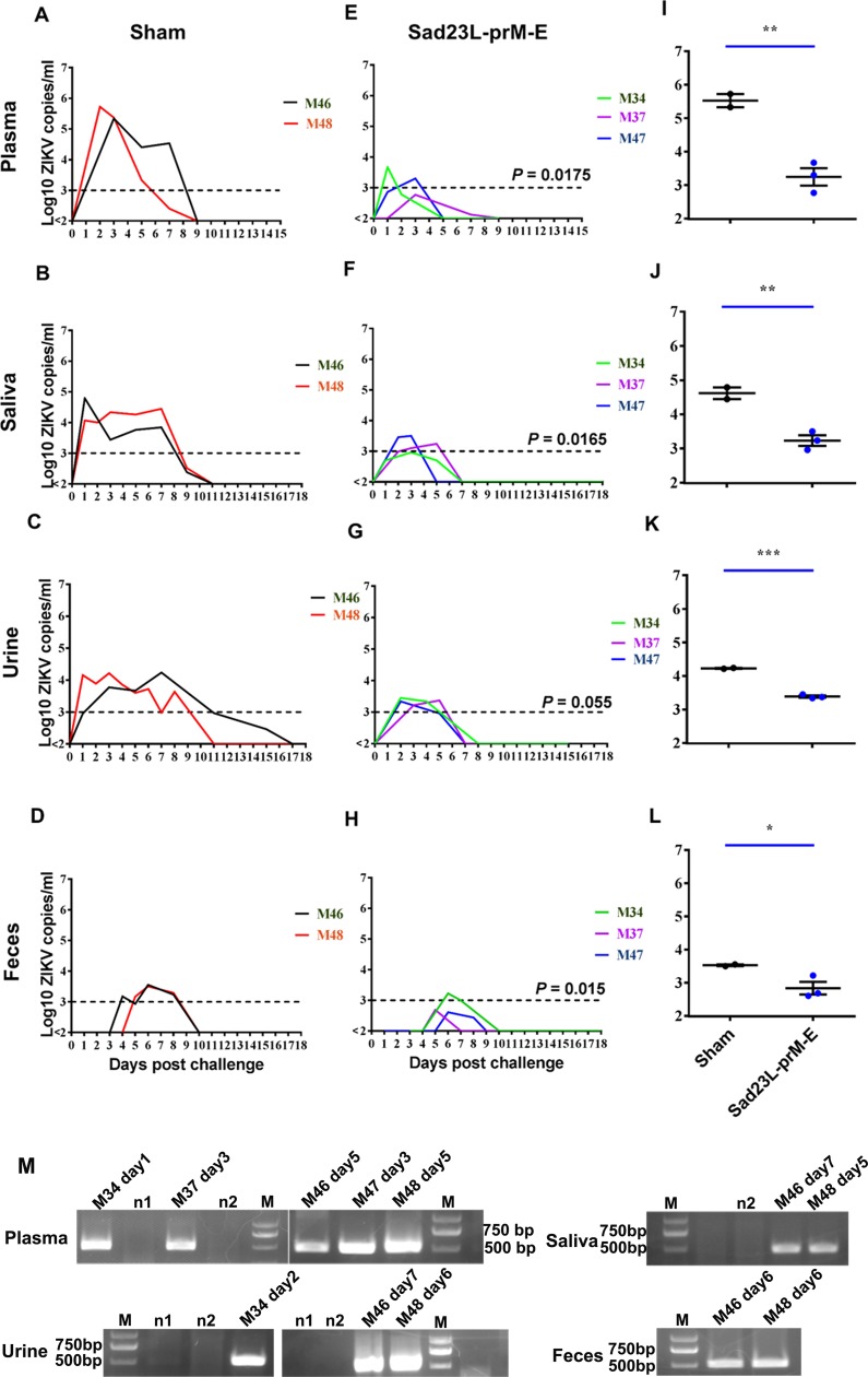
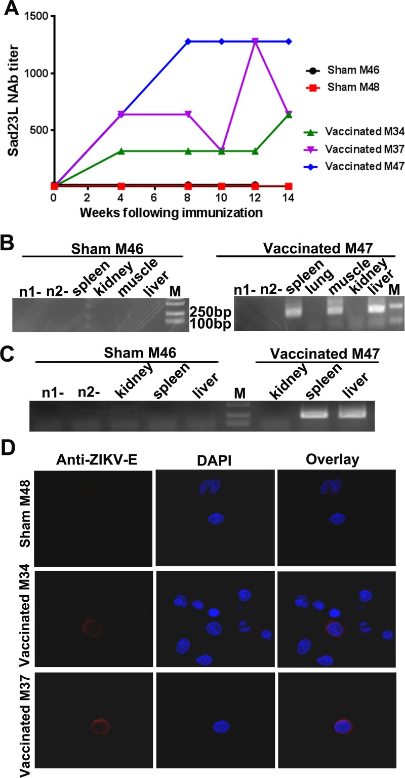
Zika virus (ZIKV) has spread in many countries or territories causing severe neurologic complications with potential fatal outcomes. The small primate common marmosets are susceptible to ZIKV, mimicking key features of human infection. Here, a novel simian adenovirus type 23 vector-based vaccine expressing ZIKV pre-membrane-envelope proteins (Sad23L-prM-E) was produced in high infectious titer. Due to determination of immunogenicity in mice, a single-dose of 3×10⁸ PFU Sad23L-prM-E vaccine was intramuscularly inoculated to marmosets. This vaccine raised antibody titers of 104.07 E-specific and 103.13 neutralizing antibody (NAb), as well as robust specific IFN-γ secreting T-cell response (1,219 SFCs/106 cells) to E peptides. The vaccinated marmosets, upon challenge with a high dose of ZIKV (105 PFU) six weeks post prime immunization, reduced viremia by more than 100 folds, and the low level of detectable viral RNA (<103 copies/ml) in blood, saliva, urine and feces was promptly eliminated when the secondary NAb (titer >103.66) and T-cell response (>726 SFCs/106 PBMCs) were acquired 1-2 weeks post exposure to ZIKV, while non-vaccinated control marmosets developed long-term high titer of ZIKV (105.73 copies/ml) (P<0.05). No significant pathological lesions were observed in marmoset tissues. Sad23L-prM-E vaccine was detectable in spleen, liver and PBMCs at least 4 months post challenge. In conclusion, a prime immunization with Sad23L-prM-E vaccine was able to protect marmosets against ZIKV infection when exposed to a high dose of ZIKV. This Sad23L-prM-E vaccine is a promising vaccine candidate for prevention of ZIKV infection in humans.


Conflict of interest statement

The authors have declared that no competing interests exist.

Figures

Fig 1
Fig 1. Characteristics of novel Sad23L-prM-E vaccine.
(A) Genomic construct of Sad23L-prM-E vaccine. Cytomegalovirus promoter (CMV), Japanese encephalitis virus signal peptide (JE signal) sequences and ZIKV prM-E genes were inserted into the deleted E1 region of simian adenovirus type 23 genome (SAdV23), the initial E3 region was deleted and E4orf6 was replaced by the corresponding element of Ad5-E4orf6. ITR indicates inverted terminal repeat sequence. (B) E protein expressions from Sad23L-prM-E virus infected naïve marmoset’s PBMCs, HEK-293, Vero and Huh7.1.5 cells were analyzed by Western blot, while Sad23L-empty virus infected cells were used as mock controls. Anti-ZIKV and anti-GAPDH antibodies were used to detect E protein and internal control protein, respectively. M indicates protein marker. (C) E protein expression in Vero cells was detected by immunofluorescence staining.
Fig 2
Fig 2. Humoral and cellular immune dose response of Sad23L-prM-E vaccine to mice.
C57BL/6 mice were immunized by a single dose of 5×10⁶, 107 or 108 PFU Sad23L-prM-E vaccine, Sad23L-empty or PBS control, respectively. Sera and splenocytes were collected from vaccinated mice for measurement of antibody and T cell responses at 4 weeks post-immunization. (A) E-Ab titer was measured by ELISA. (B) NAb titer was measured by PRNT50. (C and D) The number of specific IFN-γ spot forming cells (SFCs) per million splenocytes to M or E peptides was measured by ELISpot, respectively. (E-G) Intracellular IL-2+ CD3+CD4+ and IL-2+ CD3+CD8+ cells to M peptides were detected by ICS. (H-N) Intracellular IL-2+ CD3+CD4+, IL-2+ CD3+CD8+, IFN-γ+ CD3+CD4+ and TNF-α+ CD3+CD8+ T cells to E peptides were detected by ICS, respectively. Data are shown as mean ± SEM (standard errors of means). P values are analyzed by one-way ANOVA. Statistically significant differences are shown with asterisks (*, P<0.05; **, P< 0.01 and ***, P< 0.001); ns, no significant difference; PRNT50, 50% plaque reduction neutralization test.
Fig 3
Fig 3. Humoral and cellular immune response of common marmosets to Sad23L-prM-E vaccine.
Baseline values of antibody and T-cell responses for pre-vaccination were tested individually in the sera and PBMCs of five marmosets (week 0), and a mean value was calculated as a background for each reaction in marmosets. (A) Testing of the background of IFN-γ secreting spot forming cells (SFCs) per million PBMCs to M or E peptides by ELISpot. (B) Testing of the background of intracellular IFN-γ+ CD3+CD4+ or IFN-γ+ CD3+CD8+ cells to M or E peptides by ICS assay. (C) The baseline values of E-binding and neutralizing antibodies and T cell responses in pre-vaccination marmosets. Three marmosets were immunized with a single dose of 3×10⁸ PFU Sad23L-prM-E vaccine and two marmosets were inoculated with PBS as sham vaccine. From these vaccinated or sham marmosets, sera and PBMCs were isolated to test specific antibody and T-cell responses at 4 weeks post-immunization, and to compare with pre-vaccination values. (D) E-Ab was detected by ELISA. (E) NAb was detected with PRNT50. (F and G) The number of IFN-γ SFCs per million PBMCs to M or E peptides was measured by ELISpot, respectively. (H-J) The percent of intracellular IFN-γ+ CD3+CD4+ or IFN-γ+ CD3+CD8+ cells to M peptides was detected by ICS assay. (K-M) The percentage of intracellular IFN-γ+CD3+CD4+ or IFN-γ+CD3+CD8+ cells to E peptides was detected by ICS assay, respectively. Data are shown as a mean ± SEM (standard errors of means). P values are analyzed with one-tailed t test. Statistically significant differences are shown with asterisks (*, P<0.05; **, P< 0.01 and ***, P< 0.001). ns, no significant difference.
Fig 4
Fig 4. Protection of Sad23L-prM-E vaccine immunized marmosets against ZIKV infection.
Three marmosets (M34, M37 and M47) were immunized with a single dose of 3×10⁸ PFU Sad23L-prM-E vaccines, and two marmosets (M46 and M48) were inoculated with PBS as sham control. All animals were intramuscularly challenged with 105 PFU ZIKV in 6 weeks post vaccination. Sera and body fluids were collected daily after ZIKV challenge for determining viral loads by RT-qPCR and confirming by RT nested-PCR and sequencing. (A-D) Detection of viral load of sham marmosets from plasma, saliva, urine and feces by RT-qPCR. (E-H) Detection of viral load of vaccinated marmosets from plasma, saliva, urine and feces by RT-qPCR. (I-L) Comparison of peak viral loads between sham and vaccinated marmosets in plasma, saliva, urine and feces. (M) Confirmation of ZIKV RNA for predicted amplicons (500 bp) by RT nested-PCR and sequencing was found for plasma, saliva, urine and feces samples from sham marmosets M46 and M48 at 5th to 7th day post challenge, vaccinated M34 at 1st day in plasma and 2nd day in urine, and vaccinated M37 and M47 at 3rd day in plasma, respectively. The dashed line indicated a relatively low level of viral load. P values are analyzed by one-tailed Mann-Whitney U tests or one-tailed t test. Statistically significant differences are shown with asterisks (*, P<0.05; **, P< 0.01 and ***, P< 0.001). ns, no significant difference. n1 and n2 indicate negative control. M indicates DNA molecular markers.
Fig 5
Fig 5. Memory immune response in vaccinated marmosets against ZIKV infection over time.
Vaccinated marmosets were tested for secondary neutralizing antibody and T-cell immune response after exposure to ZIKV challenge in comparison with sham marmosets. Titration of serum neutralizing antibody from vaccinated marmosets by PRNT50; (A) in vaccinated animals, (B) in control unvaccinated animals. Measurement of IFN-γ secreting T cell response of PBMCs to E peptides by ELISpot; (C) in vaccinated marmosets, (D) in non-vaccinated sham marmosets. The number of IFN-γ spot forming cells (SFCs) per million PBMCs is calculated in ELISpot. Data are shown as a mean ± SEM (standard errors of means). Statistically significant differences of neutralizing antibody (NAb) or T cell response (E-peptides) between vaccination and sham groups are compared by one-tailed Mann-Whitney U tests, and the P values are presented in Fig 5B and 5D, respectively.
Fig 6
Fig 6. Biodistribution and expression of Sad23L-prM-E vaccine in marmosets.
(A) Neutralizing antibody titers to Sad23L vectorial virus after prime immunization with Sad23L-prM-E vaccine. (B) Nested-PCR amplifying Sad23L-hexon gene (200bp) in tissues (spleen, lung, muscle, kidney and liver) at 16 weeks after prime immunization with Sad23L-prM-E vaccine. (C) RT nested-PCR amplifying ZIKV E mRNA transcripts in tissues (spleen, muscle, kidney and liver) of marmosets at 10 weeks (72 days) post challenge. (D) Immunofluorescence staining to detect the expression of E protein in PBMCs of immunized marmosets. PBMCs were isolated from marmosets in 4 months post challenge, stained by ZIKV E antibody and DAPI. Red immunofluorescence of ZIKV E protein was detected in vaccinated marmosets M34 and M37 but not in non-vaccinated sham marmosets M48.

Similar articles

Cited by

References

    1. Dick GW, Kitchen SF, Haddow AJ. Zika virus. I. Isolations and serological specificity. Trans R Soc Trop Med Hyg. 1952; 46(5): 509–20. 10.1016/0035-9203(52)90042-4 - DOI - PubMed
    1. Zanluca C, Melo VC, Mosimann AL, Santos GI, Santos CN, et al. First report of autochthonous transmission of Zika virus in Brazil. Mem Inst Oswaldo Cruz. 2015; 110(4): 569–72. 10.1590/0074-02760150192 - DOI - PMC - PubMed
    1. Valentine G, Marquez L, Pammi M. Zika virus-associated microcephaly and eye lesions in the newborn. J Pediatric Infect Dis Soc. 2016; 5(3): 323–8. 10.1093/jpids/piw037 - DOI - PubMed
    1. Ladhani SN, O'Connor C, Kirkbride H, Brooks T, Morgan D. Outbreak of Zika virus disease in the Americas and the association with microcephaly, congenital malformations and Guillain–Barré syndrome. Arch Dis Child. 2016; 101(7): 600–2. 10.1136/archdischild-2016-310590 - DOI - PMC - PubMed
    1. Rasmussen SA, Jamieson DJ, Honein MA, Petersen LR. Zika virus and birth defects—reviewing the evidence for causality. N Engl J Med. 2016; 374(20): 1981–1987. 10.1056/NEJMsr1604338 - DOI - PubMed

Publication types

MeSH terms