Cannabinoid receptor expression in non-small cell lung cancer. Effectiveness of tetrahydrocannabinol and cannabidiol inhibiting cell proliferation and epithelial-mesenchymal transition in vitro
- PMID: 32049991
- PMCID: PMC7015420
- DOI: 10.1371/journal.pone.0228909
Cannabinoid receptor expression in non-small cell lung cancer. Effectiveness of tetrahydrocannabinol and cannabidiol inhibiting cell proliferation and epithelial-mesenchymal transition in vitro
Abstract
Background/objective: Patients with non-small cell lung cancer (NSCLC) develop resistance to antitumor agents by mechanisms that involve the epithelial-to-mesenchymal transition (EMT). This necessitates the development of new complementary drugs, e.g., cannabinoid receptors (CB1 and CB2) agonists including tetrahydrocannabinol (THC) and cannabidiol (CBD). The combined use of THC and CBD confers greater benefits, as CBD enhances the effects of THC and reduces its psychotropic activity. We assessed the relationship between the expression levels of CB1 and CB2 to the clinical features of a cohort of patients with NSCLC, and the effect of THC and CBD (individually and in combination) on proliferation, EMT and migration in vitro in A549, H460 and H1792 lung cancer cell lines.
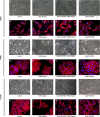
Methods: Expression levels of CB1, CB2, EGFR, CDH1, CDH2 and VIM were evaluated by quantitative reverse transcription-polymerase chain reaction. THC and CBD (10-100 μM), individually or in combination (1:1 ratio), were used for in vitro assays. Cell proliferation was determined by BrdU incorporation assay. Morphological changes in the cells were visualized by phase-contrast and fluorescence microscopy. Migration was studied by scratch recolonization induced by 20 ng/ml epidermal growth factor (EGF).
Results: The tumor samples were classified according to the level of expression of CB1, CB2, or both. Patients with high expression levels of CB1, CB2, and CB1/CB2 showed increased survival reaching significance for CB1 and CB1/CB2 (p = 0.035 and 0.025, respectively). Both cannabinoid agonists inhibited the proliferation and expression of EGFR in lung cancer cells, and CBD potentiated the effect of THC. THC and CBD alone or in combination restored the epithelial phenotype, as evidenced by increased expression of CDH1 and reduced expression of CDH2 and VIM, as well as by fluorescence analysis of cellular cytoskeleton. Finally, both cannabinoids reduced the in vitro migration of the three lung cancer cells lines used.
Conclusions: The expression levels of CB1 and CB2 have a potential use as markers of survival in patients with NSCLC. THC and CBD inhibited the proliferation and expression of EGFR in the lung cancer cells studied. Finally, the THC/CBD combination restored the epithelial phenotype in vitro.
Conflict of interest statement
The authors have declared that no competing interests exist.
Figures


Similar articles
-
In Vitro Effect of Δ9-Tetrahydrocannabinol and Cannabidiol on Cancer-Associated Fibroblasts Isolated from Lung Cancer.Int J Mol Sci. 2022 Jun 17;23(12):6766. doi: 10.3390/ijms23126766. Int J Mol Sci. 2022. PMID: 35743206 Free PMC article.
-
The diverse CB1 and CB2 receptor pharmacology of three plant cannabinoids: delta9-tetrahydrocannabinol, cannabidiol and delta9-tetrahydrocannabivarin.Br J Pharmacol. 2008 Jan;153(2):199-215. doi: 10.1038/sj.bjp.0707442. Epub 2007 Sep 10. Br J Pharmacol. 2008. PMID: 17828291 Free PMC article. Review.
-
Pharmacological data of cannabidiol- and cannabigerol-type phytocannabinoids acting on cannabinoid CB1, CB2 and CB1/CB2 heteromer receptors.Pharmacol Res. 2020 Sep;159:104940. doi: 10.1016/j.phrs.2020.104940. Epub 2020 May 26. Pharmacol Res. 2020. PMID: 32470563
-
Cannabidiol skews biased agonism at cannabinoid CB1 and CB2 receptors with smaller effect in CB1-CB2 heteroreceptor complexes.Biochem Pharmacol. 2018 Nov;157:148-158. doi: 10.1016/j.bcp.2018.08.046. Epub 2018 Sep 6. Biochem Pharmacol. 2018. PMID: 30194918
-
Delta-9-tetrahydrocannabinol/cannabidiol (Sativex®): a review of its use in patients with moderate to severe spasticity due to multiple sclerosis.Drugs. 2014 Apr;74(5):563-78. doi: 10.1007/s40265-014-0197-5. Drugs. 2014. PMID: 24671907 Review.
Cited by
-
Tetrahydrocannabinols: potential cannabimimetic agents for cancer therapy.Cancer Metastasis Rev. 2023 Sep;42(3):823-845. doi: 10.1007/s10555-023-10078-2. Epub 2023 Jan 25. Cancer Metastasis Rev. 2023. PMID: 36696005 Review.
-
Effect of Delta-9-tetrahydrocannabinol and cannabidiol on milk proteins and lipid levels in HC11 cells.PLoS One. 2022 Aug 17;17(8):e0272819. doi: 10.1371/journal.pone.0272819. eCollection 2022. PLoS One. 2022. PMID: 35976913 Free PMC article.
-
The Therapeutic Potential of the Endocannabinoid System in Age-Related Diseases.Biomedicines. 2022 Oct 6;10(10):2492. doi: 10.3390/biomedicines10102492. Biomedicines. 2022. PMID: 36289755 Free PMC article. Review.
-
Characterization of the Antitumor Potential of Extracts of Cannabis sativa Strains with High CBD Content in Human Neuroblastoma.Int J Mol Sci. 2023 Feb 14;24(4):3837. doi: 10.3390/ijms24043837. Int J Mol Sci. 2023. PMID: 36835247 Free PMC article.
-
Cannabidiol Inhibits the Proliferation and Invasiveness of Prostate Cancer Cells.J Nat Prod. 2023 Sep 22;86(9):2151-2161. doi: 10.1021/acs.jnatprod.3c00363. Epub 2023 Sep 13. J Nat Prod. 2023. PMID: 37703852 Free PMC article.
References
-
- Franklin WA, Veve R, Hirsch FR, Helfrich BA, Bunn PA Jr. Epidermal growth factor receptor family in lung cancer and premalignancy. Semin Oncol. 2002;29(1 Suppl 4):3–14. - PubMed
Publication types
MeSH terms
Substances
LinkOut - more resources
Full Text Sources
Medical
Research Materials
Miscellaneous

