Review
. 2020 Feb 10;12(2):407.
doi: 10.3390/cancers12020407.

Multiple Myeloma: Available Therapies and Causes of Drug Resistance

Affiliations
Review

Multiple Myeloma: Available Therapies and Causes of Drug Resistance

Vanessa Pinto et al. Cancers (Basel). .

Abstract

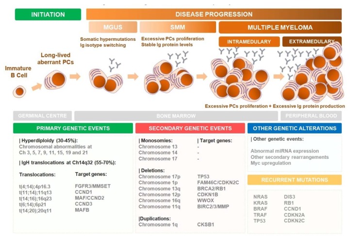
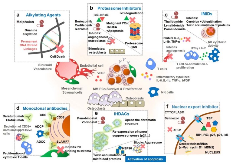
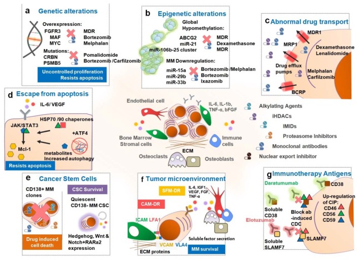
Multiple myeloma (MM) is the second most common blood cancer. Treatments for MM include corticosteroids, alkylating agents, anthracyclines, proteasome inhibitors, immunomodulatory drugs, histone deacetylase inhibitors and monoclonal antibodies. Survival outcomes have improved substantially due to the introduction of many of these drugs allied with their rational use. Nonetheless, MM patients successively relapse after one or more treatment regimens or become refractory, mostly due to drug resistance. This review focuses on the main drugs used in MM treatment and on causes of drug resistance, including cytogenetic, genetic and epigenetic alterations, abnormal drug transport and metabolism, dysregulation of apoptosis, autophagy activation and other intracellular signaling pathways, the presence of cancer stem cells, and the tumor microenvironment. Furthermore, we highlight the areas that need to be further clarified in an attempt to identify novel therapeutic targets to counteract drug resistance in MM patients.

Keywords: drug resistance; drug response; multiple myeloma; treatment.


Conflict of interest statement

R.B.: Grants and research fund: Celgene, AMGEN/SPH/APCL; Advisory boards—AMGEN, Celgene, Janssen and Takeda; Speaker honoraria—AMGEN, Celgene, Janssen and Takeda. J.E.G.: Speaker’s bureau—Abbvie, Janssen, Pfizer, Roche; Advisory boards—Abbvie, Pfizer, Roche. M.H.V., R.B. and H.C. are members of the research team of a project financed by Celgene and M.H.V. is member of the team of a grant co-financed by AMGEN. These companies had no role in the decision to publish nor were they involved in the writing of this manuscript. The authors declare no conflict of interest.

Figures

Figure 1
Figure 1
Biology of multiple myeloma (MM) development and progression. MM is the end stage of a multistep neoplastic transformation of PCs. Virtually all MM initiates as a monoclonal gammopathy of undetermined significance (MGUS). In this early stage, a BM PC may accumulate several primary genetic mutations (as chromosomal abnormalities and/or IgH translocations) that affect the expression of key target genes (e.g., cyclins, FGFR3, MYC deregulation, etc.) granting a proliferative advantage to these mutated cells. The presence of abnormal amounts of PCs in the BM with slightly increased levels of Ig proteins but no clinical symptoms is defined as smoldering multiple myeloma (SMM). Nevertheless, these hyperproliferative PCs will endure additional secondary genetic mutations that aggravate this aberrant phenotype leading to the accumulation of high amounts of PCs in the BM and consequently to the secretion of excessive levels of Igs towards the blood stream. Ultimately, this will lead to the clinical manifestation of severe symptoms (as hypercalcemia, renal insufficiency, anemia and bone lesions) that define MM. This disease may progress to extramedullary disease in more advanced stages. Additional aberrant genetic events, such as mutations, deletions, methylations and microRNA (miRNA) abnormalities may occur during MM development defining the aggressiveness of the disease and response to therapy.
Figure 2
Figure 2
Therapeutic options to counteract MM. Treatments used for symptomatic or active MM include corticosteroids, alkylating agents, proteasome inhibitors (PIs), immunomodulatory drugs (IMIDs), monoclonal antibodies (mAbs), histone deacetylase inhibitors (iHDACs) and nuclear export inhibitors. (a) Classical alkylating agents as melphalan target highly proliferating cells, including malignant PCs, by intercalating permanently their DNA, causing cell death later on. (b) PIs as bortezomib, carfilzomib and ixazomib block the IkB and/or pro-apoptotic proteins degradation in malignant plasma cells proteosome, overcoming their resistance to apoptotic stimuli. (c) IMIDs as thalidomide, lenalidomide and pomalidomide modulate the inflammatory environment of the BM inhibiting the progression of MM [e.g., reduction of Interleukin-6 (IL-6), tumor necrosis factor-α (TNF-α), etc.] through inhibition of angiogenesis and other key stromal-MM cell interactions). Some of these drugs target the cereblon protein of the E3 ubiquitin ligase complex blocking the ubiquitination process in malignant PCs. This in turn leads to a toxic accumulation of proteins and cell death. (d) Monoclonal antibodies (mAbs) as daratumumab, isatuximab and elotuzumab bind to specific antigens on the surface of malignant PCs. This will in turn induce MM plasma cell death by antibody-dependent cell-mediated cytotoxicity (ADCC), complement-dependent cytotoxicity (CDC) and/or antibody-dependent cellular phagocytosis (ADCP). (e) histone deacetylase inhibitors (iHDACs) such as panobinostat and vorinostat act on malignant PCs by opening the chromatin structure. Consequently, this will activate the expression of tumor suppressor genes, which were previously silenced by aberrant histone acetylation in malignant PCs. (f) Exportin 1 (XP01) inhibitors as selinexor act on malignant PCs by blocking the export tumor suppressor proteins out of the nucleus by the XPO1 pump while retaining many oncoprotein mRNAs within the nucleus.
Figure 3
Figure 3
Mechanisms of Drug Resistance enforced by MM. (a) Genetic alterations such as t(4;14), t(14;16) and t(14;20) translocations, 17p and 13p deletions and c-Myc associated abnormalities are associated with an unfavorable prognosis and insufficient response to current treatments. Additionally, (b) epigenetic alterations induced by a global hypomethylation of the DNA leads to the abnormal expression of several genes such as ATP binding cassette super-family G member 2 (ABCG2) and several miRs (e.g., miR-21, -15a, -29b, etc.) in malignant plasma cells (PCs) conferring a multidrug resistance (MDR) phenotype. (c) The overexpression of drug efflux pumps, namely P-glycoprotein (P-gp), in the malignant PCs mediates the cellular efflux of several drugs lowering intracellular drug concentration to sub-lethal levels. Moreover, (d) alterations in NF-κB, phosphatidylinositol 3-kinase/ protein kinase B (PI3K/AKT), mitogen-activated protein kinase/ extracellular signal-regulated kinase (MAPK/ERK) and Janus kinase/signal transducer and activator of transcription 3 (JAK/STAT3) signaling pathways in these PCs confers resistance to apoptotic stimuli and evasion to drug-induced cell death. (e) CD138- MM putative cancer stem cells are intrinsically resistant to most drugs. This small subset of cancer stem cells (CSCs) will survive therapy and remain as undetected/quiescent residual disease. Later on, these CSCs have the ability to self-initiate MM and cause refractory post-treatment relapse. (f) The bone marrow microenvironment is essential for MM survival, development and drug resistance by secretion of soluble factors e.g., interleukin 6 (IL-6), insulin-like growth factor 1 (IGF-1), vascular endothelial growth factor (VEGF), B-cell activating factor (BAFF), fibroblast growth factor (FGF), stromal cell-derived factor 1α (SDF1α), and tumor necrosis factor-α (TNF-α). (g) Immunotherapy antigens: the anti-CD38 and SLAM7 monoclonal antibodies (mAbs), daratumumab and elotuzumab, fail to reach therapeutic efficacy either due to extracellular binding of the mAbs to target antigens or to upregulation of cell surface expression of the complement-inhibitors proteins CD46, CD56 and CD59.

References

    1. Kumar S.K., Rajkumar V., Kyle R.A., van Duin M., Sonneveld P., Mateos M.-V., Gay F., Anderson K.C. Multiple myeloma. Nat. Rev. Dis. Primers. 2017;3:nrdp201746. doi: 10.1038/nrdp.2017.46. - DOI - PubMed
    1. Kumar S., Paiva B., Anderson K.C., Durie B., Landgren O., Moreau P., Munshi N., Lonial S., Bladé J., Mateos M.-V., et al. International Myeloma Working Group consensus criteria for response and minimal residual disease assessment in multiple myeloma. Lancet Oncol. 2016;17:e328–e346. doi: 10.1016/S1470-2045(16)30206-6. - DOI - PubMed
    1. Kyle R.A., Gertz M.A., Witzig T.E., Lust J.A., Lacy M.Q., Dispenzieri A., Fonseca R., Rajkumar V.S., Offord J.R., Larson D.R., et al. Review of 1027 Patients with Newly Diagnosed Multiple Myeloma. Mayo Clin. Proc. 2003;78:21–33. doi: 10.4065/78.1.21. - DOI - PubMed
    1. Landgren O., Graubard B., Katzmann J., Kyle R., Ahmadizadeh I., Clark R., Kumar S., Dispenzieri A., Greenberg A., Therneau T., et al. Racial disparities in the prevalence of monoclonal gammopathies: A population-based study of 12 482 persons from the National Health and Nutritional Examination Survey. Leukemia. 2014;28:1537–1542. doi: 10.1038/leu.2014.34. - DOI - PMC - PubMed
    1. Kumar S., Dispenzieri A., Lacy M., Gertz M., Buadi F., Pandey S., Kapoor P., Dingli D., Hayman S., Leung N., et al. Continued improvement in survival in multiple myeloma: Changes in early mortality and outcomes in older patients. Leukemia. 2014;28:1122–1128. doi: 10.1038/leu.2013.313. - DOI - PMC - PubMed

LinkOut - more resources