. 2020 Feb 12;22(1):24.
doi: 10.1186/s13075-020-2110-9.

Tumour necrosis factor alpha promotes secretion of 14-3-3η by inducing necroptosis in macrophages

Affiliations

Tumour necrosis factor alpha promotes secretion of 14-3-3η by inducing necroptosis in macrophages

Gulzhan Trimova et al. Arthritis Res Ther. .

Abstract

Background: 14-3-3η is an intracellular protein also detected in the serum and synovial fluid of patients with rheumatoid arthritis (RA). It is closely related to disease activity and anti-cyclic citrullinated peptide antibody levels. However, the main source of 14-3-3η and the mechanism of its release into the extracellular space remain unclear. Addressing these two points was the main goal of the current study.

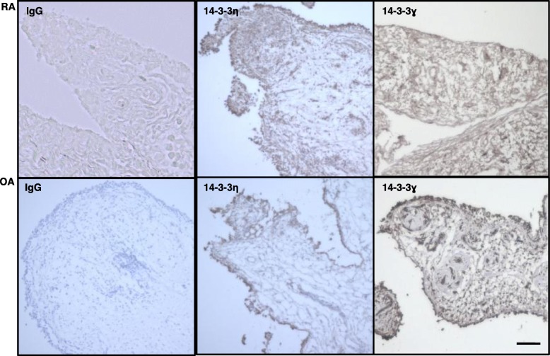
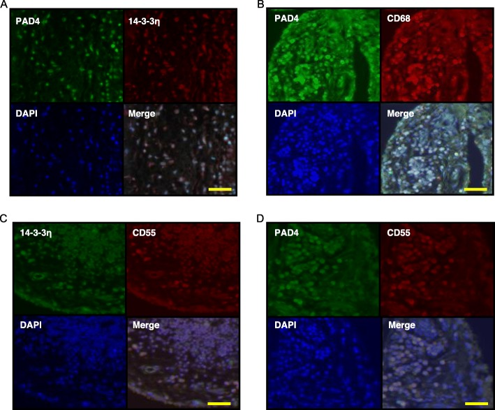
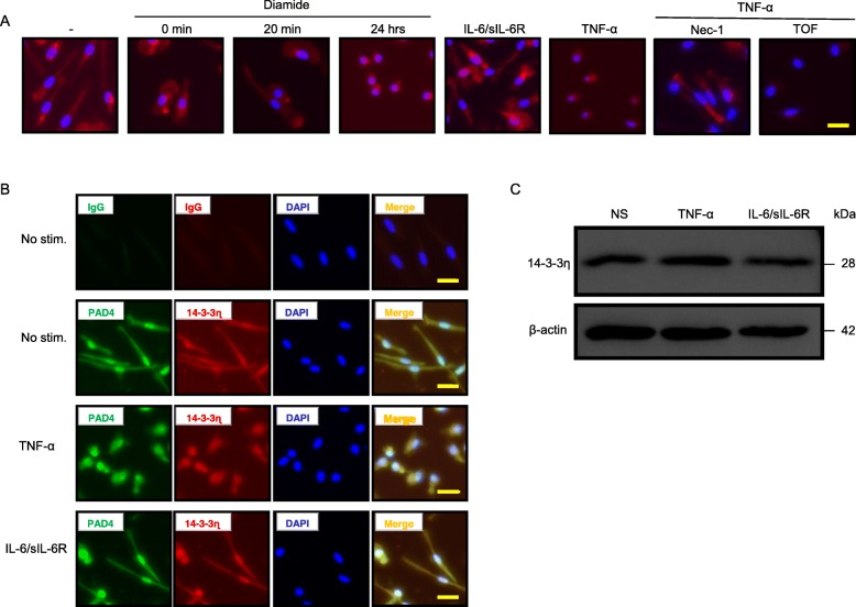
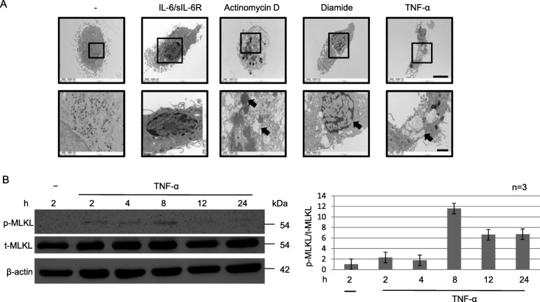
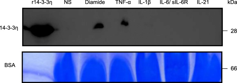
Methods: The source of 14-3-3η was investigated by immunostaining RA synovial tissue. Fibroblast-like synoviocytes, CD4+ cells, and macrophages were selected as candidates among the various cell types in the synovial tissue. Phosphorylation of mixed-lineage kinase domain-like pseudokinase (MLKL) and cell death of macrophages were studied by phalloidin staining and electron microscopy after stimulation with an oxidative stress inducer (diamide) or tumour necrosis factor (TNF)-α. Extracellular 14-3-3η protein levels were examined by western blotting.

Results: Macrophages from the synovial tissue from RA, but not osteoarthritis, showed dense and widespread cytoplasmic staining for the 14-3-3η protein, co-localized with peptidylarginine deiminase 4. Swelling and membrane rupture of macrophages were induced by treatment with TNF-α, but not interleukin (IL) 6/soluble IL-6 receptor (sIL-6R). Increased MLKL phosphorylation followed by necroptosis was also induced in TNF-α-stimulated macrophages. Necrostatin-1, a necroptosis inhibitor, antagonized MLKL phosphorylation. High levels of 14-3-3η were detected in the culture supernatants of macrophages stimulated with diamide and TNF-α, but not IL-6/sIL-6R.

Conclusions: Macrophages that highly express 14-3-3η undergo TNF-α-induced necroptosis with damage to the cellular structure, resulting in the secretion of 14-3-3η into the extracellular space. The current study provides a novel mechanism for 14-3-3η level increase in the RA synovial fluid.

Keywords: 14-3-3η; Macrophage; Necroptosis; Rheumatoid arthritis; TNF-α.


Conflict of interest statement

YT has received consulting fees, speaking fees, and/or honoraria from Daiichi-Sankyo, Astellas, Pfizer, Mitsubishi-Tanabe, Bristol-Myers, Chugai, YL Biologics, Eli Lilly, Sanofi, Janssen, and UCB, and has received research grants from Mitsubishi-Tanabe, Takeda, Bristol-Myers, Chugai, Astellas, Abbvie, MSD, Daiichi-Sankyo, Pfizer, Kyowa-Kirin, Eisai, and Ono. SN has received speaking fees from Bristol-Myers, UCB, Astellas, Abbvie, Eisai, Pfizer, and Takeda, and has received research grants from Mitsubishi-Tanabe, Novartis, and MSD. KY has received research grants from Mitsubishi-Tanabe. SH has received speaking fee and honoraria from AbbVie, Asahi-Kasei Pharma, Astellas, Ayumi, Bristol-Myers Squibb, Celgene, Chugai, Eisai, Eli Lilly, Janssen, Kissei, Novartis, Pfizer, Sanofi, Takeda, Tanabe-Mitsubishi, and UCB. NB is an employee of Augurex. All other authors declare that they have no competing interests.

Figures

Fig. 1
Fig. 1
Higher expression of 14-3-3η+ and fewer 14-3-3ɣ+ cells were detectable in the RA synovium than in OA synovium. Synovium specimens of patients with RA (n = 3) or OA (n = 3) were incubated with the indicated primary antibodies and then visualized using DAB chromogen. Representative images from three independent experiments are shown. Panels labelled IgG, isotype control. Scale bar, 50 μm
Fig. 2
Fig. 2
14-3-3η is expressed in CD68+ cells but not in CD4+ T cells from the synovium of RA patients. Synovium specimens of patients with RA (n = 3) were analysed by IF using specific antibodies against 14-3-3η and CD68 (a) or CD4 (b). c Lung tissue specimens of patients with RA (n = 3) were analysed using specific antibodies against 14-3-3η and CD68. Representative images of three independent experiments are shown. Scale bar, 50 μm
Fig. 3
Fig. 3
PAD4 is localized in CD68+ cells and CD55+ cells in the RA synovium. Synovium (n = 3) specimens of patients with RA were assessed by IF using primary antibodies against PAD4 and 14-3-3η (a), PAD4 and CD68 (b), 14-3-3η and CD55 (c), or PAD4 and CD55 (d) and by FITC or rhodamine-labelled secondary antibodies. Representative images from three independent experiments are shown. Scale bar, 50 μm
Fig. 4
Fig. 4
TNF-α induces abnormal cell morphology of macrophages from HD. Macrophages were cultured with or without diamide (100 nM; for the indicated time; n = 3), IL-6/sIL-6R (10 ng/ml; 24 h; n = 3), and TNF-α (10 ng/ml; 24 h; n = 3) in the presence or absence of nec-1 (20 nM; n = 3) or TOF (300 nM; n = 3). The cells were then stained with specific antibodies against phalloidin (a) or isotype control, PAD4, or 14-3-3η (b), and DAPI (a, b). c 14-3-3η levels were detected by WB. Representative images from three independent experiments are shown. Scale bar, 50 μm
Fig. 5
Fig. 5
TNF-α induces cell death of macrophages from HD. a Macrophages were cultured with or without IL-6/sIL-6R (10 ng/ml; n = 3), actinomycin D (1 μg/ml; n = 3), diamide (1 mM, n = 3), or TNF-α (100 ng/ml; n = 3) for 24 h and then analysed by TEM. b WCLs prepared from macrophages cultured with or without TNF-α (100 ng/ml; n = 3) were analysed by IB using specific antibodies against the total or phosphorylation form of MLKL or β-actin. Quantification data for representative images from three independent experiments (n = 3) are shown. Scale bar, 5 μm (upper panel), 2 μm (lower panel)
Fig. 6
Fig. 6
14-3-3η is detectable in culture supernatants of macrophages derived from HD and treated with TNF-α. The culture supernatants of macrophages cultured with or without diamide, TNF-α (100 ng/ml; n = 3), IL-1β (10 ng/ml; n = 3), IL-6/sIL-6R (10 ng/ml; n = 3), or IL-21 (10 ng/ml; n = 3) were subjected to WB. Recombinant 14-3-3η was used as a positive control. BSA was used as a loading control and was stained with CBB-R350. Representative images from three independent experiments are shown

References

    1. McInnes IB, Schett G. Pathogenetic insights from the treatment of rheumatoid arthritis. Lancet. 2017;389:2328–2337. doi: 10.1016/S0140-6736(17)31472-1. - DOI - PubMed
    1. Smolen JS, Aletaha D, McInnes IB. Rheumatoid arthritis. Lancet. 2016;388:2023–2038. doi: 10.1016/S0140-6736(16)30173-8. - DOI - PubMed
    1. Schett G, Elewaut D, McInnes IB, Dayer JM, Neurath MF. How cytokine networks fuel inflammation: toward a cytokine-based disease taxonomy. Nat Med. 2013;19:822–824. doi: 10.1038/nm.3260. - DOI - PubMed
    1. Tanaka Y. Intensive treatment and treatment holiday of TNF-inhibitors in rheumatoid arthritis. Curr Opin Rheumatol. 2012;24:319–326. doi: 10.1097/BOR.0b013e3283524e4c. - DOI - PubMed
    1. Tanaka Y. Clinical immunity in bone and joints. J Bone Miner Metabolism. 2019;37:2–8. doi: 10.1007/s00774-018-0965-5. - DOI - PubMed

Publication types

MeSH terms