Randomized Controlled Trial
. 2020 Jul;69(7):1518-1527.
doi: 10.2337/db19-0869. Epub 2020 Feb 12.

Novel Detection and Restorative Levodopa Treatment for Preclinical Diabetic Retinopathy

Affiliations
Randomized Controlled Trial

Novel Detection and Restorative Levodopa Treatment for Preclinical Diabetic Retinopathy

Cara T Motz et al. Diabetes. 2020 Jul.

Abstract

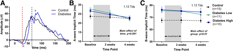
Diabetic retinopathy (DR) is diagnosed clinically by directly viewing retinal vascular changes during ophthalmoscopy or through fundus photographs. However, electroretinography (ERG) studies in humans and rodents have revealed that retinal dysfunction is demonstrable prior to the development of visible vascular defects. Specifically, delays in dark-adapted ERG oscillatory potential (OP) implicit times in response to dim-flash stimuli (<-1.8 log cd · s/m2) occur prior to clinically recognized DR. Animal studies suggest that retinal dopamine deficiency underlies these early functional deficits. In this study, we randomized individuals with diabetes, without clinically detectable retinopathy, to treatment with either low- or high-dose Sinemet (levodopa plus carbidopa) for 2 weeks and compared their ERG findings with those of control subjects (no diabetes). We assessed dim-flash-stimulated OP delays using a novel handheld ERG system (RETeval) at baseline and 2 and 4 weeks. RETeval recordings identified significant OP implicit time delays in individuals with diabetes without retinopathy compared with age-matched control subjects (P < 0.001). After 2 weeks of Sinemet treatment, OP implicit times were restored to control values, and these improvements persisted even after a 2-week washout. We conclude that detection of dim-flash OP delays could provide early detection of DR and that Sinemet treatment may reverse retinal dysfunction.

Trial registration: ClinicalTrials.gov NCT02706977.


Figures

Figure 1
Figure 1
This waveform is an averaged representation of the ERG recordings and measurements to dim (A and B) and bright (C and D) flash stimuli from the control group. The a- and b-waves were marked first. OPs were filtered offline and then overlaid onto the full ERG waveform. OP1 was identified as the first peak after the a-wave trough. Red asterisks indicate the trough and peak of each OP wave.
Figure 2
Figure 2
Dim-flash stimuli revealed ERG delays in diabetic eyes without retinopathy. A: OP2 delays in participants with diabetes were significantly delayed compared with control subjects in response to dim-flash stimuli (1.13 Tds; P < 0.01). B: In contrast, bright-flash stimuli (85 Tds) did not reveal any differences in OP implicit times in the same subject groups. Dim-flash stimuli also revealed delays in a-waves (P < 0.05) and b-waves (P < 0.001) in eyes from the group with diabetes compared with control subjects (C and E), while a- and b-wave implicit times to bright-flash stimuli (D and F) were similar. *P < 0.05, **P < 0.01, ***P < 0.001.
Figure 3
Figure 3
Sinemet treatments improve OP implicit times in eyes in the group with diabetes without retinopathy. A: After 2 days of low-dose treatment, inner retinal function, as measured by the OP implicit time, was significantly improved (Student paired t test: t = 4.54, P = 0.001). High-dose treatment produced faster OP2 implicit times in the majority of eyes in the group with diabetes but did not reach statistical significance. B: After 2 weeks of Sinemet treatments, both high- and low-dose groups had OP2 implicit times that were indistinguishable from control subjects. This effect was maintained at 4 weeks, following a 2-week washout period of the drug. C: Representative OP waveforms from a Diabetes High participant at baseline and 4 weeks (2 weeks of Sinemet treatment plus 2-week washout period). The OP waveforms are overlaid with the full ERG. Red asterisks indicate the OP peaks and show the improvement in implicit time across all OPs. Arrowheads indicate OP2 peaks.
Figure 4
Figure 4
Sinemet treatment did not change OP timing in response to bright-flash stimuli or in eyes that did not have a delay. A: In response to bright flash, Sinemet treatment did not alter the OP implicit time. B: Eyes from participants with diabetes who had normal OP implicit times were not affected by the treatment. These results indicate that Sinemet only benefited eyes in which rod-driven inner retinal function was abnormal.
Figure 5
Figure 5
High- and low-dose Sinemet treatment resulted in faster a-wave implicit times in eyes from the group with diabetes. A: Representative control and diabetic waveforms that illustrate the delay in a- and b-wave at baseline. The red and gray dashed vertical lines indicate the a- and b-wave peaks in the control waveform, respectively. The gray arrowheads indicate the delayed peak for each wave. B: The a-wave implicit times were significantly improved at 2 weeks with values that became similar to the control group by 4 weeks. C: The b-wave implicit times were slightly improved by 4 weeks but did not reach control values.
Figure 6
Figure 6
Visual function did not change in participants with diabetes and control participants with Sinemet treatment. A: Visual acuity thresholds were not different between the diabetic and control eyes at baseline. Sinemet did not alter thresholds. B: Contrast sensitivity function for diabetic and control eyes was identical with a peak at 2 c/d. C: Plot of contrast sensitivity at 2.0 c/d across time shows that treatment did not alter contrast sensitivity thresholds. a.u., arbitrary units.

Comment in

References

    1. Cho NH, Shaw JE, Karuranga S, et al. . IDF Diabetes Atlas: global estimates of diabetes prevalence for 2017 and projections for 2045. Diabetes Res Clin Pract 2018;138:271–281 - PubMed
    1. Stefánsson E, Bek T, Porta M, Larsen N, Kristinsson JK, Agardh E. Screening and prevention of diabetic blindness. Acta Ophthalmol Scand 2000;78:374–385 - PubMed
    1. Frank RN. Diabetic retinopathy. N Engl J Med 2004;350:48–58 - PubMed
    1. Sharma S, Oliver-Fernandez A, Liu W, Buchholz P, Walt J. The impact of diabetic retinopathy on health-related quality of life. Curr Opin Ophthalmol 2005;16:155–159 - PubMed
    1. Gross JG, Glassman AR, Liu D, et al. .; Diabetic Retinopathy Clinical Research Network . Five-year outcomes of panretinal photocoagulation vs intravitreous ranibizumab for proliferative diabetic retinopathy: a randomized clinical trial [published correction appears in JAMA Ophthalmol 2019;137:467]. JAMA Ophthalmol 2018;136:1138–1148 - PMC - PubMed

Publication types

Associated data