. 2020 Mar 3;117(9):5074-5080.
doi: 10.1073/pnas.1913463117. Epub 2020 Feb 12.

A three-dimensional hybrid electrode with electroactive microbes for efficient electrogenesis and chemical synthesis

Affiliations

A three-dimensional hybrid electrode with electroactive microbes for efficient electrogenesis and chemical synthesis

Xin Fang et al. Proc Natl Acad Sci U S A. .

Abstract

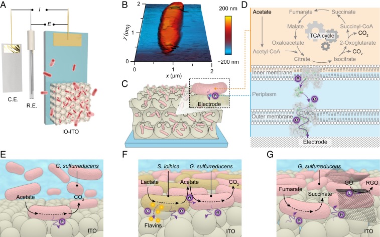
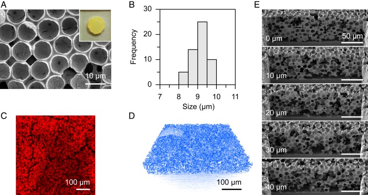
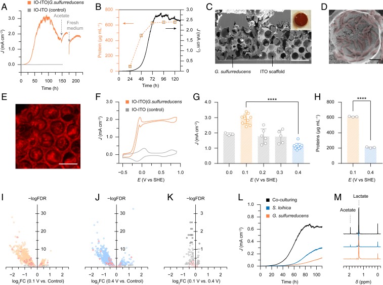
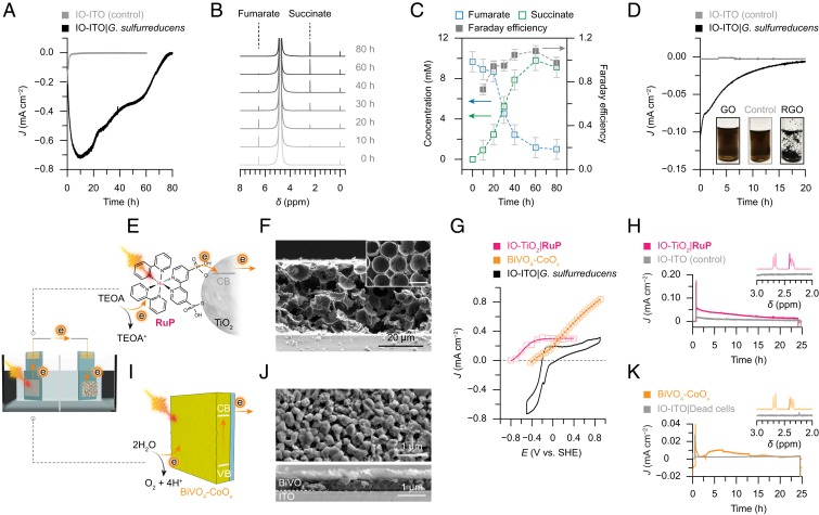
Integration of electroactive bacteria into electrodes combines strengths of intracellular biochemistry with electrochemistry for energy conversion and chemical synthesis. However, such biohybrid systems are often plagued with suboptimal electrodes, which limits the incorporation and productivity of the bacterial colony. Here, we show that an inverse opal-indium tin oxide electrode hosts a large population of current-producing Geobacter and attains a current density of 3 mA cm-2 stemming from bacterial respiration. Differential gene expression analysis revealed Geobacter's transcriptional regulations to express more electron-relaying proteins when interfaced with electrodes. The electrode also allows coculturing with Shewanella for syntrophic electrogenesis, which grants the system additional flexibility in converting electron donors. The biohybrid electrode containing Geobacter can also catalyze the reduction of soluble fumarate and heterogenous graphene oxide, with electrons from an external power source or an irradiated photoanode. This biohybrid electrode represents a platform to employ live cells for sustainable power generation and biosynthesis.

Keywords: Geobacter; electrogenesis; electrosynthesis.


Conflict of interest statement

The authors declare no competing interest.

Figures

Fig. 1.
Fig. 1.
Schematic representation of microbial electrogenesis and electrosynthesis within the IO-ITO electrodes. (A) An IO-ITO|G. sulfurreducens electrode is assembled into a three-electrode system with a counter electrode (C.E.) and a reference electrode (R.E.). (B) Atomic force microscopy (AFM) image of G. sulfurreducens on a silicon wafer. (C) Schematic representation of a biohybrid electrode where G. sulfurreducens colonized on the IO-ITO scaffold. (D) Extracellular electron transfer at the interface between G. sulfurreducens and an electrode. Acetate is metabolized into CO2 via the TCA cycle and excess electrons are discharged to an external electrode via OMCs. (E) Schematic representation of microbial electrogenesis. G. sulfurreducens is respiring on an electrode surface with acetate as the electron donor while continuously releasing electrons to the electrode. (F) Syntrophic electrogenesis where S. loihica metabolizes lactate into acetate and transfers electrons to the electrode mainly through self-excreted flavins. G. sulfurreducens then consumes acetate and releases electrons to the electrode. (G) Microbial electrosynthesis of succinate and RGO using a biohybrid IO-ITO electrode. At negative potentials, the sessile G. sulfurreducens exploits exogenously supplied electrons to maintain its metabolism while transferring excess reducing equivalent to soluble fumarate and heterogeneous GO.
Fig. 2.
Fig. 2.
Structure of the IO-ITO electrode. (A) Top-view SEM image. Inset shows a photograph of the electrode (S = 0.25 cm2). (B) Histogram of the pore size distribution of the IO-ITO electrode. (C) CLSM image of the IO-ITO electrode, showing channels that allow bacteria to penetrate. A total of 20 μL of rhodamine B solution (5 mM, in methanol) was dropcast on an IO-ITO electrode and dried in the dark. Excitation: 552 nm. Emission: 590 to 640 nm. (D) X-ray microscopy image of the interconnected IO-ITO scaffold (colored in blue, see Movie S1). (E) Serial cross-sectional SEM images of the IO-ITO electrode acquired from FIB-SEM. Cross-sectional views of every 10 μm are displayed (Movie S2).
Fig. 3.
Fig. 3.
IO-ITO electrodes as the platform to accommodate electroactive bacteria for microbial electrogenesis. (A) A representative current of G. sulfurreducens respiring inside an IO-ITO electrode at 0.1 V vs. SHE with acetate (40 mM, pH 7.4). A bare IO-ITO electrode was used as a control. The two arrows indicate the addition of 40 mM acetate to the existing medium and the replenishing of a fresh medium containing 40 mM acetate, respectively. (B) Colorimetric quantification of proteins in the hybrid electrodes during bacterial colonization at 0.1 V vs. SHE (a typical current output shown as the black trace). (C) Cross-sectional SEM image of an IO-ITO|G. sulfurreducens electrode. (Scale bar: 20 μm.) The Inset shows a photograph of the electrode (S = 0.25 cm2). (D) G. sulfurreducens (artificially colored in red) attached on the surface of an IO-ITO electrode. (Scale bar: 2 μm.) (E) CLSM image of an IO-ITO|G. sulfurreducens electrode. The hybrid electrodes were stained with 5-cyano-2,3-ditolyl tetrazolium chloride (CTC, 10 mM) and incubated in the dark for 30 min at 25 °C. Excitation: λex = 488 nm, emission: λem = 600 to 650 nm. (F) Representative CV scans of an IO-ITO|G. sulfurreducens electrode and a bare IO-ITO electrode (control) with acetate. The redox wave near 0 V vs. SHE is derived from the medium solution. Scan rate: 5 mV s−1. (G) Potential dependence of the current produced by IO-ITO|G. sulfurreducens. Independent samples: 0.0 V: n = 3; 0.1 V: n = 10; 0.2 V: n = 6; 0.3 V: n = 6; 0.4 V: n = 10. (H) Colorimetric quantification of proteins in IO-ITO|G. sulfurreducens electrodes prepared at 0.1 V and 0.4 V vs. SHE. n = 3 independent samples. Error bars represent the standard error of the mean. Significance value: ****P < 0.0001. (IK) Volcano plots of differential gene expression of G. sulfurreducens in IO-ITO electrodes at 0.1 V and 0.4 V vs. SHE. (I) 0.1 V vs. control; (J) 0.4 V vs. control; (K) 0.1 V vs. 0.4 V. The control group was the planktonic G. sulfurreducens anaerobically cultured in a medium solution with 20 mM acetate and 50 mM fumarate (pH 7.2) at 30 °C. The expression difference is represented by the log fold change in base 2 (log2FC) versus a baseline group (I and J, control; K, 0.4 V). The expression difference is considered significant provided that the false discovery rate (FDR), the adjusted P value for multiple testing, is less than 0.05 (−logFDR > 1.3). Positive logFC values represent higher expression compared with the baseline group. The red points indicate the genes encoding putative c-type cytochromes in G. sulfurreducens identified by ref. . Each point represents the average value of one transcript in three replicates. (L) Representative currents of G. sulfurreducens, S. loihica, and a mixed culture of G. sulfurreducens and S. loihica, with an IO-ITO electrode at 0.4 V vs. SHE with lactate (40 mM, pH 7.4). (M) 1H NMR spectra of the electrolyte solution extracted after 100 h electrogenesis with IO-ITO|G. sulfurreducens, IO-ITO|S. loihica, and IO-ITO|mixed cultures. TMSP-d4 (1 mM) was used as the reference (0 ppm) and internal standard for quantification. 1H NMR peaks of acetate (singlet, 1.92 ppm) and lactate (doublet, 1.34 ppm) are indicated. All of the electrochemical experiments were performed under a N2:CO2 atmosphere (80:20, v:v%) at 30 °C.
Fig. 4.
Fig. 4.
Microbial electrosynthesis and photoelectrosynthesis with IO-ITO|G. sulfurreducens electrodes. (A) Representative cathodic current of an IO-ITO|G. sulfurreducens electrode catalyzing fumarate (10 mM, pH 7.4) reduction at −0.45 V vs. SHE. A bare IO-ITO electrode was used as a control. (B) 1H NMR spectra of the electrolyte solution aliquoted during the course of reaction. TMSP-d4 (1 mM) was used as the reference (0 ppm) and internal standard for quantification. 1H NMR peaks of fumarate (singlet, 6.52 ppm) and succinate (singlet, 2.41 ppm) are indicated. (C) Quantification of reactants and products and Faraday efficiency during the course of reaction. (D) Cathodic current of an IO-ITO|G. sulfurreducens electrode reducing GO (0.1 mg mL−1) at −0.3 V vs. SHE. A bare IO-ITO electrode was used as a control. The Inset shows photographs of GO solutions before (labeled “GO”) and after reduction by a bare IO-ITO (labeled “Control”) and an IO-ITO|G. sulfurreducens electrode (labeled “RGO”). All of the reactions were performed in a N2:CO2 atmosphere (80:20 v:v%) at 30 °C, with Pt and Ag/AgCl as counter and reference electrode, respectively. (E) Schematic representation of a PEC cell consisting of an IO-TiO2|RuP anode and an IO-ITO|G. sulfurreducens cathode. Under irradiation, the excited RuP* dye injects an electron into the conduction band of the TiO2 electrode, which is further directed to the cathode via an external circuit. The RuP+ dye is regenerated by extracting an electron from TEOA. (F) SEM image of an IO-TiO2 electrode. The Inset shows the top view of the electrode (Scale bar: 10 µm.) The IO-TiO2 electrode has a thickness of 40 µm and macropore size of 10 µm. (G) Photocurrent from chronoamperometry of the IO-TiO2|RuP (0.25 cm2) and BiVO4-CoOx (1.0 cm2) photoanodes (plotted at different applied potentials) and cyclic voltammogram of the IO-ITO|G. sulfurreducens electrode in fumarate (10 mM, pH 7.2) solution. Three-electrode configuration, scan rate: 5 mV s−1. (H) Light-driven fumarate reduction with an IO-TiO2|RuP||IO-ITO|G. sulfurreducens two-electrode system at zero bias. A bare IO-ITO electrode without bacteria was used as the cathode for a control experiment (gray trace). TEOA (25 mM, in 50 mM KCl) was used as the electron donor for the photoanode. (I) Schematic representation of a PEC cell consisting of a BiVO4-CoOx anode and an IO-ITO|G. sulfurreducens cathode. BiVO4 absorbs light and donates excited electrons to the external circuit while oxidizing water with the aid of the CoOx cocatalyst. (J) Top-view (Top) and cross-sectional (Bottom) SEM images of a BiVO4-CoOx electrode. The thickness of BiVO4 film was 500 nm and CoOx cocatalysts were deposited on top. (K) Light-driven fumarate reduction with a BiVO4-CoOx||IO-ITO|G. sulfurreducens two-electrode system at zero bias. A hybrid electrode with dead bacteria (deactivated by 0.1% glutaraldehyde) was used as the cathode for a control experiment (gray trace). A PBS solution (20 mM Na2HPO4, pH 7.3) was used for the photoanode compartment. The Insets in H and K are 1H NMR spectra of the solution extracted from the cathode compartment after 24 h of irradiation. TMSP-d4 (1 mM) was used as the reference (0 ppm) and internal standard for quantification. The NMR peak of succinate (singlet, 2.41 ppm) is highlighted and the doublet peak at 2.7 ppm is assigned to malate. Conditions: 20 mM fumarate, pH 7.2, U = 0 V, I = 100 mW cm−2, AM 1.5G, in a N2:CO2 (80:20 v:v%) atmosphere at 25 °C. The photocurrent was normalized to the geometrical area of the cathode (0.25 cm2).

References

    1. Kornienko N., Zhang J. Z., Sakimoto K. K., Yang P., Reisner E., Interfacing nature’s catalytic machinery with synthetic materials for semi-artificial photosynthesis. Nat. Nanotechnol. 13, 890–899 (2018). - PubMed
    1. Sakimoto K. K., Kornienko N., Yang P., Cyborgian material design for solar fuel production: The emerging photosynthetic biohybrid systems. Acc. Chem. Res. 50, 476–481 (2017). - PubMed
    1. Reetz M. T., Biocatalysis in organic chemistry and biotechnology: Past, present, and future. J. Am. Chem. Soc. 135, 12480–12496 (2013). - PubMed
    1. Lee J. W., et al. , Systems metabolic engineering of microorganisms for natural and non-natural chemicals. Nat. Chem. Biol. 8, 536–546 (2012). - PubMed
    1. Smanski M. J., et al. , Synthetic biology to access and expand nature’s chemical diversity. Nat. Rev. Microbiol. 14, 135–149 (2016). - PMC - PubMed

Publication types

LinkOut - more resources