. 2020 Mar 31;94(8):e01910-19.
doi: 10.1128/JVI.01910-19. Print 2020 Mar 31.

Mutational Functional Analysis of the Pseudorabies Virus Nuclear Egress Complex-Nucleocapsid Interaction

Affiliations

Mutational Functional Analysis of the Pseudorabies Virus Nuclear Egress Complex-Nucleocapsid Interaction

Sebastian Rönfeldt et al. J Virol. .

Abstract

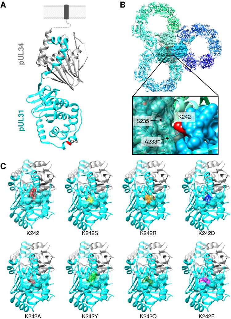
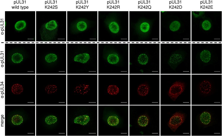
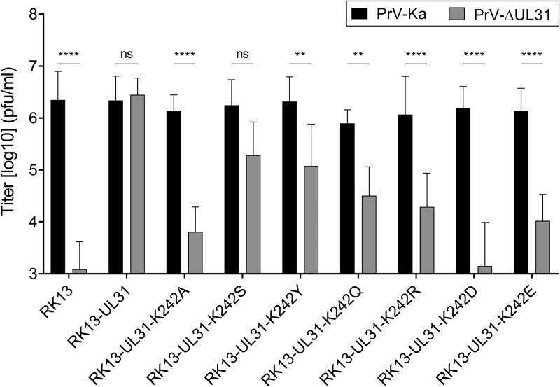
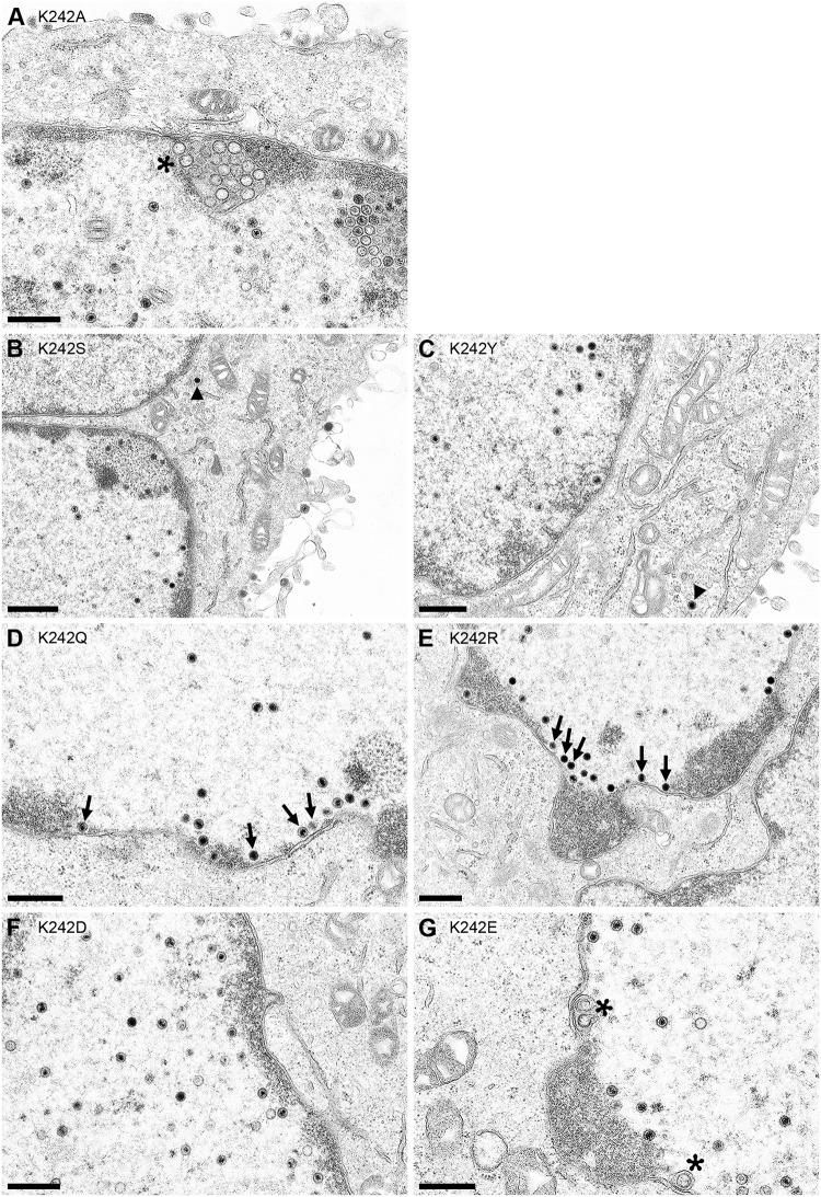
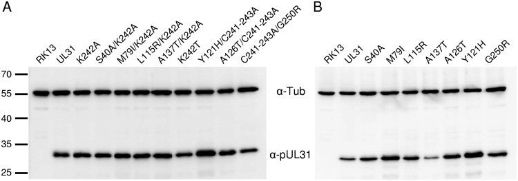
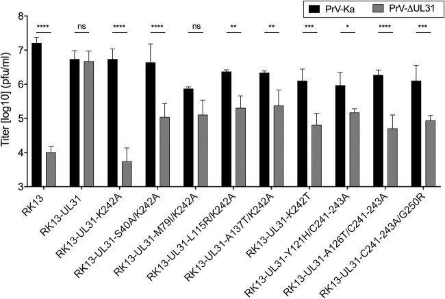
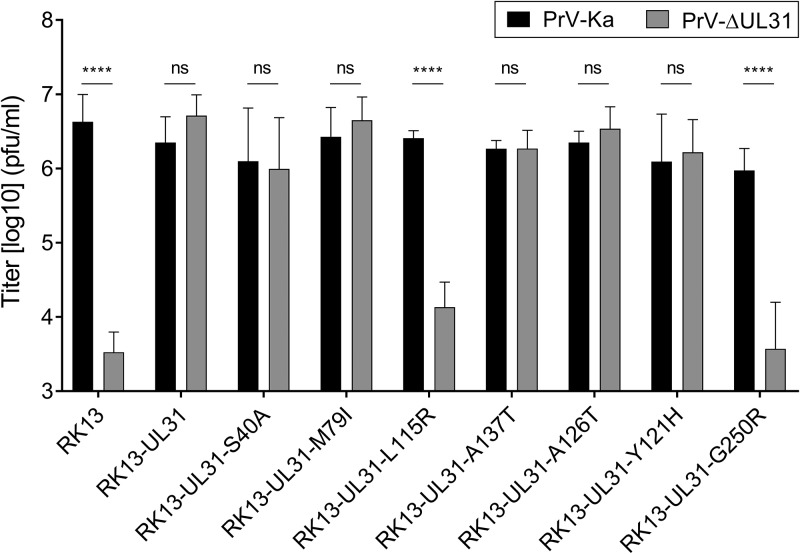
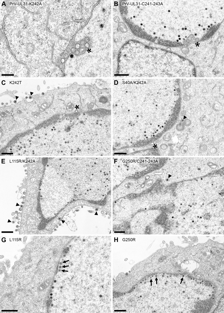
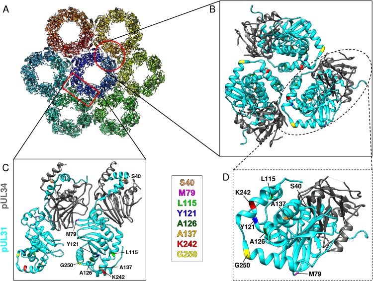
Herpesvirus nucleocapsids leave the nucleus by a vesicle-mediated translocation mediated by the viral nuclear egress complex (NEC). The NEC is composed of two conserved viral proteins, designated pUL34 and pUL31 in the alphaherpesvirus pseudorabies virus (PrV). It is required for efficient nuclear egress and is sufficient for vesicle formation and scission from the inner nuclear membrane (INM). Structure-based mutagenesis identified a lysine at position 242 (K242) in pUL31, located in the most membrane distal part of the NEC, to be crucial for efficient nucleocapsid incorporation into budding vesicles. Replacing the lysine by alanine (K242A) resulted in accumulations of empty vesicles in the perinuclear space, despite the presence of excess nucleocapsids in the nucleus. However, it remained unclear whether the defect in capsid incorporation was due to interference with a direct, electrostatic interaction between the capsid and the NEC or structural restrictions. To test this, we replaced K242 with several amino acids, thereby modifying the charge, size, and side chain orientation. In addition, virus recombinants expressing pUL31-K242A were passaged and screened for second-site mutations. Compensatory mutations at different locations in pUL31 or pUL34 were identified, pointing to an inherent flexibility of the NEC. In summary, our data suggest that the amino acid at position 242 does not directly interact with the nucleocapsid but that rearrangements in the NEC coat are required for efficient nucleocapsid envelopment at the INM.IMPORTANCE Herpesviruses encode an exceptional vesicle formation and scission machinery, which operates at the inner nuclear membrane, translocating the viral nucleocapsid from the nucleus into the perinuclear space. The conserved herpesviral nuclear egress complex (NEC) orchestrates this process. High-resolution imaging approaches as well as the recently solved crystal structures of the NEC provided deep insight into the molecular details of vesicle formation and scission. Nevertheless, the molecular mechanism of nucleocapsid incorporation remained unclear. In accordance with structure-based predictions, a basic amino acid could be pinpointed in the most membrane-distal domain of the NEC (pUL31-K242), indicating that capsid incorporation might depend on a direct electrostatic interaction. Our follow-up study, described here, however, shows that the positive charge is not relevant but that the overall structure matters.

Keywords: herpesvirus; nuclear egress complex; nuclear envelope; pUL31; pUL34; pseudorabies virus.


Figures

FIG 1
FIG 1
Structural prediction of the PrV NEC. (A) pUL34 is shown in gray and pUL31 is shown in cyan in the PrV NEC structure (17). The orientation toward and anchorage in the INM are indicated by the dotted line, and the transmembrane domain in a lipid bilayer is represented by a dark gray box. The location of the amino acid (K242) targeted in this study is indicated in red. (B) Bottom view of the membrane-distal end in the honeycomb array (17), with interacting interhexameric molecules being shown by surface representation with amino acid K242 in red. The zoomed image of the boxed region shows the interaction interface of different NEC heterodimers with one pUL31 in a semitransparent surface presentation, while the neighboring pUL31 is shown with its full surface. Amino acids in close proximity to K242 are indicated by arrows and given with the corresponding position. (C) A bottom view of the membrane-distal end of pUL31 with a partial surface presentation of alpha helix H10 is shown with amino acid K242 in red. Amino acid substitutions at position 242 are modeled into the NEC dimer structure (PDB accession number 5E8C). Molecular graphics and analysis were performed with the UCSF Chimera package (34).
FIG 2
FIG 2
Intracellular localization of pUL31 mutants and colocalization with pUL34. Localization was tested after transfection of RK13 cells with the corresponding pUL31 expression plasmids (top row). Colocalization was analyzed after cotransfection with pcDNA-UL34. pUL31 was stained with a pUL31-specific monoclonal antibody (green, second row), while for pUL34, polyclonal anti-pUL34 serum (red, third row) was used. Merged channels are shown in the bottom row. Fluorescence was imaged with a confocal laser-scanning microscope (63× oil immersion objective, single slice; SP5; Leica Germany). Bars, 10 μm.
FIG 3
FIG 3
pUL31 expression in RK13 cells. Lysates of cell lines stably expressing native or mutated pUL31 as well as nontransgenic RK13 cells were separated in an SDS-10% polyacrylamide gel. Proteins were detected after transfer to nitrocellulose with the monospecific anti-pUL31 rabbit serum. As loading control, anti-α-tubulin was used. The molecular masses of the marker proteins (in kilodaltons) are indicated on the left.
FIG 4
FIG 4
trans-Complementation assays. Functional complementation by the generated pUL31 mutants was tested after infection of the stably expressing cell lines with PrV-Ka and PrV-ΔUL31 at an MOI of 5. Cells and supernatant were harvested at 24 h p.i., and titers were determined on RK13-UL31 cells. Shown are the mean values from four independent experiments with the corresponding standard deviations. Statistically significant differences are indicated (**, P ≤ 0.01; ****, P ≤ 0.0001; ns, not significant).
FIG 5
FIG 5
Ultrastructural analyses of cells expressing pUL31 mutants infected with PrV-ΔUL31. RK13-UL31-K242A (A), RK13-UL31-K242S (B), RK13-UL31-K242Y (C), RK13-UL31-K242Q (D), RK13-UL31-K242R (E), RK13-UL31-K242D (F), and RK13-UL31 K242E (G) cells were infected with PrV-ΔUL31 at an MOI of 1 and processed for electron microscopy at 14 h p.i. Representative images are shown. Asterisks mark empty vesicles in the PNS, arrows point to nucleocapsids close to the INM, and arrowheads indicate nucleocapsids or virions in the cytoplasm. Bars, 500 nm (A, C to G) and 800 nm (B).
FIG 6
FIG 6
Immunoblot analysis of RK13 cells expressing pUL31 with second-site mutations. RK13 cells expressing UL31-K242A with the second-site mutations (A) or pUL31 carrying only these mutations (B) were harvested and tested for pUL31 expression with the monospecific anti-pUL31 serum. Anti-α-tubulin (α-Tub) was used as a loading control.
FIG 7
FIG 7
Complementation of PrV-ΔUL31 by second-site-mutated pUL31-K242A and pUL31-C241-243A. Complementation by pUL31-K242A and pUL31-C241-243A carrying second-site mutations was tested after infection of the stably expressing cell lines with PrV-Ka and PrV-ΔUL31 at an MOI of 5. Cells and supernatant were harvested at 24 h p.i., and titers were determined on RK13-UL31 cells. Shown are the mean values from three independent experiments with the corresponding standard deviations. Statistically significant differences were evaluated using GraphPad Prism software and are indicated (*, P ≤ 0.05; **, P ≤ 0.01; ***, P ≤ 0.001; ****, P ≤ 0.0001; ns, not significant).
FIG 8
FIG 8
Effect of second-site mutations in the absence of the K242A mutation or the C241-243A mutations. The K242A and C241-243A mutations in plasmids expressing the pUL31 genes derived from the passaged mutants were repaired to the wild type by site-directed mutagenesis. Stably expressing cells were infected with PrV-Ka and PrV-ΔUL31 as described in the Fig. 7 legend. Shown are mean values from three independent experiments with the corresponding standard deviations (****, P ≤ 0.0001; ns, not significant).
FIG 9
FIG 9
Ultrastructural analysis. RK13 cells were infected with PrV-UL31-K242A (A) or PrV-UL31-C241-243A (B). (C to H) RK13 cells expressing pUL31-K242T (C), pUL31-S40A/K242A (D), pUL31-L115R/K242A (E), pUL31-G250R/C241-243A (F), pUL31-L115R (G), or pUL31-G250R (H), infected with PrV-ΔUL31 (MOI, 1), fixed, and processed for electron microscopy at 14 h p.i. Asterisks mark empty vesicles in the PNS, nucleocapsids close to the INM are indicated by arrows, and nucleocapsids or virions in the cytoplasm or at the plasma membrane are highlighted by arrowheads. Bars, 300 nm (A) and 600 nm (B to H).
FIG 10
FIG 10
Location of the pUL31 mutations in the NEC dimer and in the hexagonal lattice. (A) Top view of the NEC hexameric lattice (PDB accession number 5FKI) (17, 21). The chains are rainbow colored. (B to D) Close-up views of NEC heterodimer interfaces of the hexameric lattice, with the mutations being marked in color. pUL31 is shown in cyan, while pUL34 is shown in gray. The interfaces indicated in panel A are represented by a red circle for panel B and a red rectangle for panel C. (B) Top view of the trimer interface between hexamers. (C) Side view of the dimer-dimer interface. (D) Zoom of the membrane-distal part of the NEC shown in panel B. The amino acids that were mutated are highlighted by different colors and labeled. Molecular graphics and analysis were performed with the UCSF Chimera package (34).

Similar articles

Cited by

  • Nuclear Egress.
    Draganova EB, Thorsen MK, Heldwein EE. Draganova EB, et al. Curr Issues Mol Biol. 2021;41:125-170. doi: 10.21775/cimb.041.125. Epub 2020 Aug 7. Curr Issues Mol Biol. 2021. PMID: 32764158 Free PMC article. Review.
  • Alphaherpesvirus in Pets and Livestock.
    Duan SH, Li ZM, Yu XJ, Li D. Duan SH, et al. Microorganisms. 2025 Jan 4;13(1):82. doi: 10.3390/microorganisms13010082. Microorganisms. 2025. PMID: 39858850 Free PMC article. Review.

References

    1. Johnson DC, Baines JD. 2011. Herpesviruses remodel host membranes for virus egress. Nat Rev Microbiol 9:382–394. doi:10.1038/nrmicro2559. - DOI - PubMed
    1. Mettenleiter TC, Klupp BG, Granzow H. 2009. Herpesvirus assembly: an update. Virus Res 143:222–234. doi:10.1016/j.virusres.2009.03.018. - DOI - PubMed
    1. Mettenleiter TC, Muller F, Granzow H, Klupp BG. 2013. The way out: what we know and do not know about herpesvirus nuclear egress. Cell Microbiol 15:170–178. doi:10.1111/cmi.12044. - DOI - PubMed
    1. Mettenleiter TC. 2002. Herpesvirus assembly and egress. J Virol 76:1537–1547. doi:10.1128/jvi.76.4.1537-1547.2002. - DOI - PMC - PubMed
    1. Bigalke JM, Heuser T, Nicastro D, Heldwein EE. 2014. Membrane deformation and scission by the HSV-1 nuclear egress complex. Nat Commun 5:4131. doi:10.1038/ncomms5131. - DOI - PMC - PubMed

Publication types

MeSH terms

LinkOut - more resources