

Challenging the concept that eumelanin is the polymorphic brown banded pigment in Cepaea nemoralis
- PMID: 32051478
- PMCID: PMC7016172
- DOI: 10.1038/s41598-020-59185-y
Challenging the concept that eumelanin is the polymorphic brown banded pigment in Cepaea nemoralis
Abstract
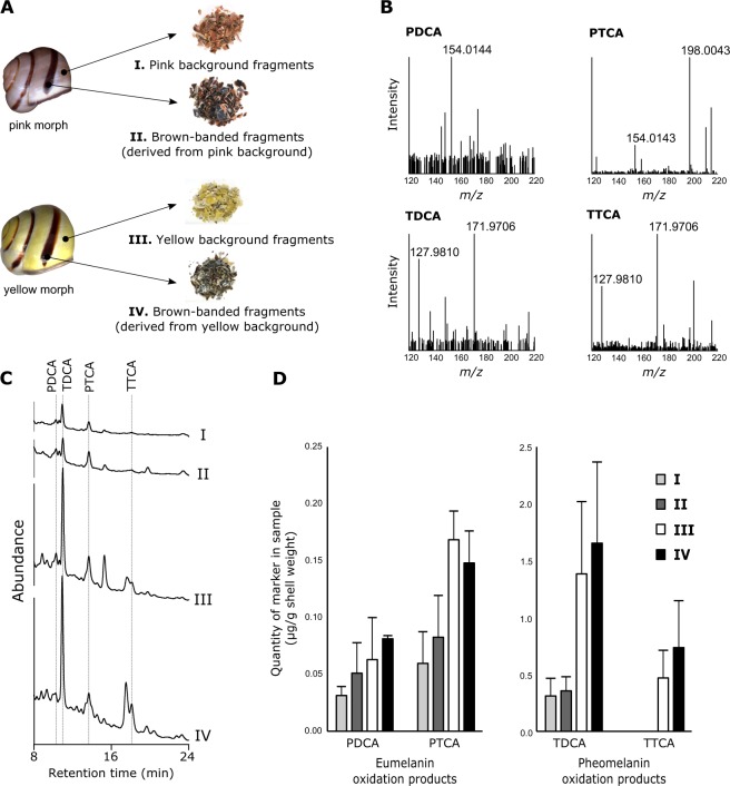
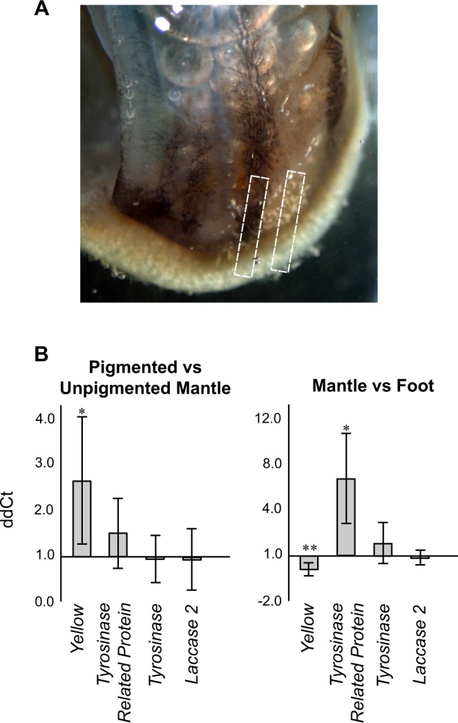
The common grove snail Cepaea nemoralis displays a stable pigmentation polymorphism in its shell that has held the attention of scientists for decades. While the details of the molecular mechanisms that generate and maintain this diversity remain elusive, it has long been employed as a model system to address questions related to ecology, population genetics and evolution. In order to contribute to the ongoing efforts to identify the genes that generate this polymorphism we have tested the long-standing assumption that melanin is the pigment that comprises the dark-brown bands. Surprisingly, using a newly established analytical chemical method, we find no evidence that eumelanin is differentially distributed within the shells of C. nemoralis. Furthermore, genes known to be responsible for melanin deposition in other metazoans are not differentially expressed within the shell-forming mantle tissue of C. nemoralis. These results have implications for the continuing search for the supergene that generates the various pigmentation morphotypes.
Conflict of interest statement
The authors declare no competing interests.
Figures
References
-
- Fisher RA, Diver C. Crossing-over in the Land Snail Cepaea nemoralis, L. Nature. 1934;133:834. doi: 10.1038/133834b0. - DOI
-
- Clarke B, Diver C, Murray J. Studies on Cepaea VI. The spatial and temporal distribution of pheno-types in a colony of Cepaea nemoralis (L.) Philos. Trans. R. Soc. Lond. B. Biol. Sci. 1968;253:519.
-
- Cook LM. A two-stage model for Cepaea polymorphism. Proc. R. Soc. Lond. B. Biol. Sci. 1997;353:1577. doi: 10.1098/rstb.1998.0311. - DOI
-
- Ożgo M, Schilthuizen M. Evolutionary change in Cepaea nemoralis shell colour over 43 years. Glob. Change Biol. 2012;18:74. doi: 10.1111/j.1365-2486.2011.02514.x. - DOI
Publication types
MeSH terms
Substances
LinkOut - more resources
Full Text Sources
Other Literature Sources
Research Materials
