

Is tau in the absence of amyloid on the Alzheimer's continuum?: A study of discordant PET positivity
- PMID: 32051933
- PMCID: PMC7001143
- DOI: 10.1093/braincomms/fcz046
Is tau in the absence of amyloid on the Alzheimer's continuum?: A study of discordant PET positivity
Abstract
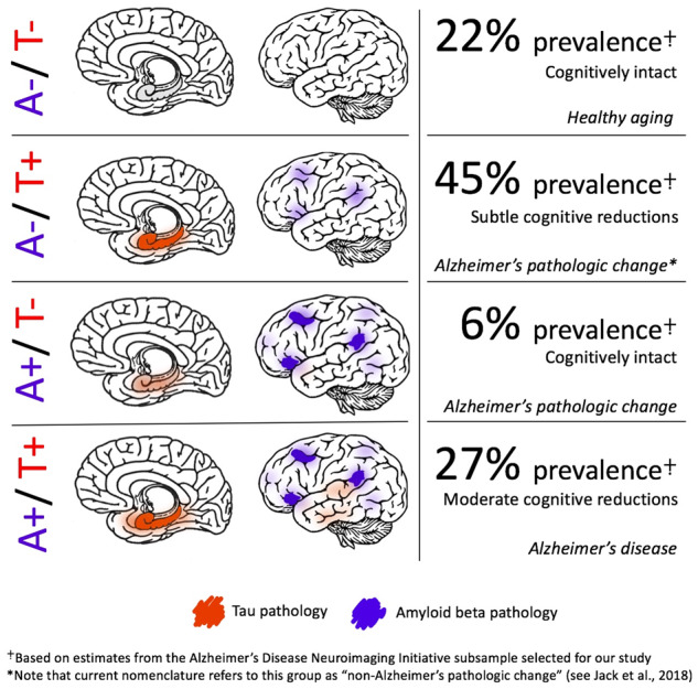
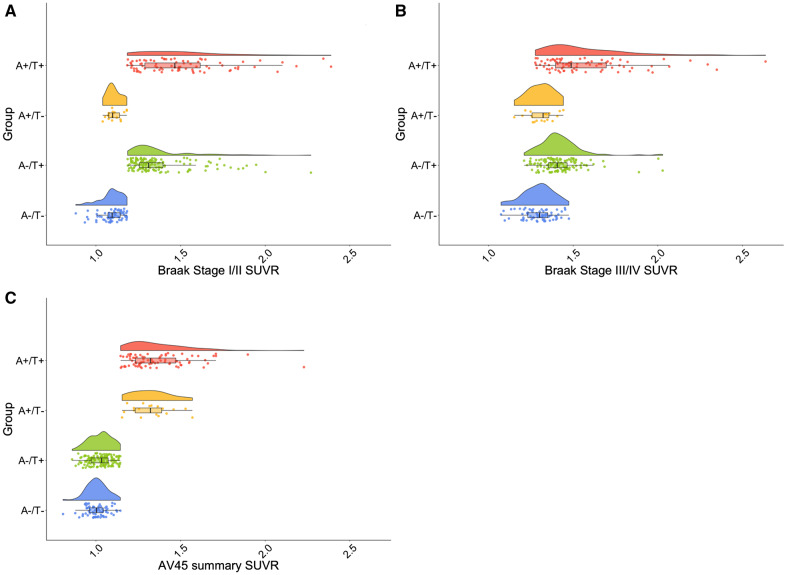
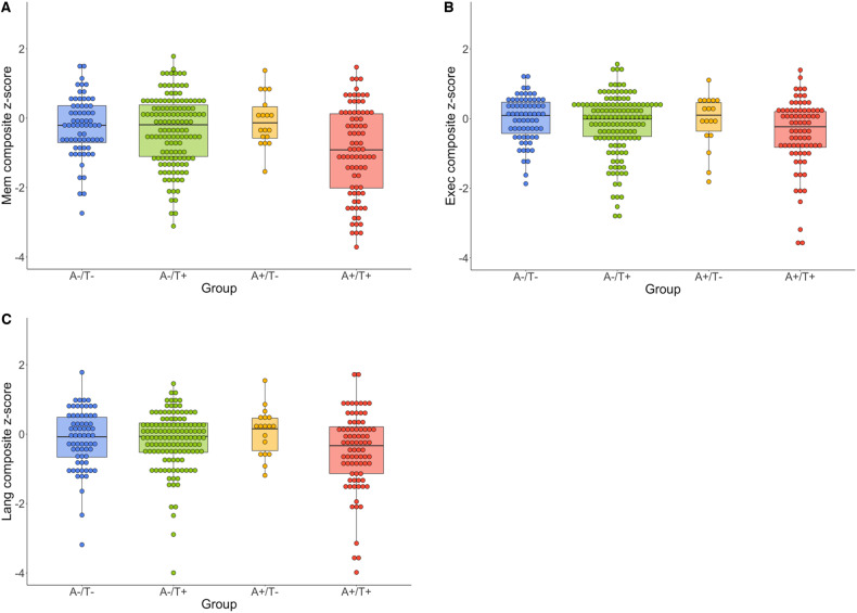
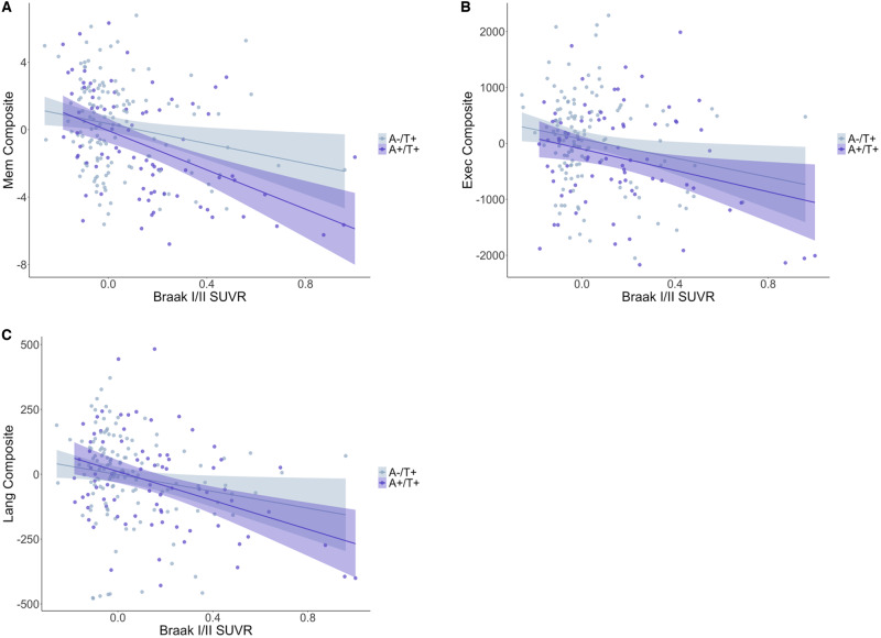
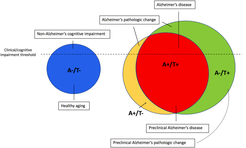
The amyloid cascade model of Alzheimer's disease posits the primacy of amyloid beta deposition preceding tau-mediated neurofibrillary tangle formation. The amyloid-tau-neurodegeneration biomarker-only diagnostic framework similarly requires the presence of amyloid beta for a diagnosis on the Alzheimer's continuum. However, medial temporal lobe tau pathology in the absence of amyloid beta is frequently observed at autopsy in cognitively normal individuals, a phenomenon that may reflect a consequence of aging and has been labelled 'primary age-related tauopathy'. Alternatively, others argue that this tauopathy reflects an early stage of the developmental continuum leading to Alzheimer's disease. We used positron emission tomography imaging to investigate amyloid beta and tau positivity and associations with cognition to better inform the conceptualization of biomarker changes in Alzheimer's pathogenesis. Five hundred twenty-three individuals from the Alzheimer's Disease Neuroimaging Initiative who had undergone flortaucipir positron emission tomography imaging were selected to derive positron emission tomography positivity thresholds using conditional inference decision tree regression. A subsample of 301 individuals without dementia (i.e. those with normal cognition or mild cognitive impairment) had also undergone florbetapir positron emission tomography imaging within 12 months and were categorized into one of the four groups based on cortical amyloid and Braak stage I/II tau positivity: A-/T-, A+/T-, A-/T+, or A+/T+. Tau positivity in the absence of amyloid beta positivity (i.e. A-/T+) comprised the largest group, representing 45% of the sample. In contrast, only 6% of the sample was identified as A+/T-, and the remainder of the sample fell into A-/T- (22%) or A+/T+ (27%) categories. A-/T- and A+/T- groups had the best cognitive performances across memory, language and executive function; the A-/T+ group showed small-to-moderate relative decreases in cognition; and the A+/T+ group had the worst cognitive performances. Furthermore, there were negative associations between Braak stage I/II tau values and all cognitive domains only in the A-/T+ and A+/T+ groups, with strongest associations for the A+/T+ group. Among our sample of older adults across the Alzheimer's pathological spectrum, 7-fold fewer individuals have positron emission tomography evidence of amyloid beta pathology in the absence of tau pathology than the converse, challenging prevailing models of amyloid beta's primacy in Alzheimer's pathogenesis. Given that cognitive performance in the A-/T+ group was poorer than in individuals without either pathology, our results suggest that medial temporal lobe tau without cortical amyloid beta may reflect an early stage on the Alzheimer's pathological continuum.
Keywords: Alzheimer’s disease; amyloid imaging; biomarkers; mild cognitive impairment; tau imaging.
© The Author(s) (2019). Published by Oxford University Press on behalf of the Guarantors of Brain.
Figures
References
-
- Alzheimer A. Über eine eigenartige Erkrankung der Hirnrinde. Allg Zeitschr Psychiatr Psychiatr Gerichtl Med 1907; 146–8.
-
- Alzheimer’s Disease Neuroimaging Initiative; 2019. http://adni.loni.usc.edu/ (15 July 2019, date last accessed).
-
- Arriagada PV, Marzloff K, Hyman BT.. Distribution of Alzheimer-type pathologic changes in nondemented elderly individuals matches the pattern in Alzheimer's disease. Neurology 1992; 42: 1681–8. - PubMed
Grants and funding
LinkOut - more resources
Full Text Sources
Other Literature Sources