. 2020 Feb 12;61(1):4.
doi: 10.1186/s40529-019-0280-z.

Ultrastructural changes during the symbiotic seed germination of Gastrodia elata with fungi, with emphasis on the fungal colonization region

Affiliations

Ultrastructural changes during the symbiotic seed germination of Gastrodia elata with fungi, with emphasis on the fungal colonization region

Yuan-Yuan Li et al. Bot Stud. .

Abstract

Background: Gastrodia elata is a fully mycoheterotrophic orchid and has long been used in traditional Chinese medicine. The life cycle of G. elata requires an association with two different fungi-Mycena for seed germination and Armillaria for tuber growth. The association with Armillaria is representative of the phytophagous type of orchid mycorrhiza: the intracellular hyphae are lysed without forming condensed pelotons. However, whether the association with Mycena during seed germination belongs to the same type of orchid mycorrhiza is unknown.

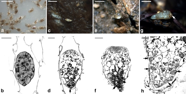
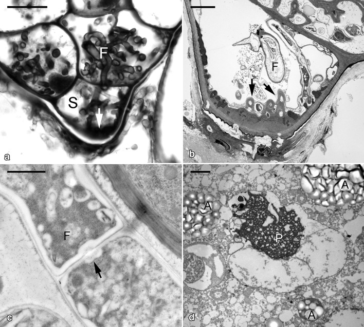
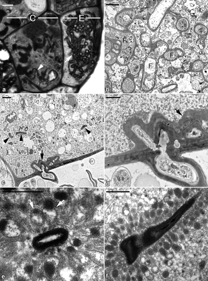
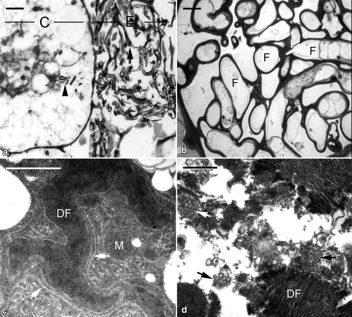
Results: Histological and ultrastructural studies revealed several notable features in different developmental stages. First, a thickened cell wall with papillae-like structures appeared during fungal penetration in the suspensor end cell, epidermal cells and cortical cells of germinating embryos. In addition, the formation of two distinctive cell types in the colonized region of a protocorm (i.e., the passage canal cell filled with actively growing fungal hyphae) can be observed in the epidermal cell, and the distinctive digestion cell with a dense cytoplasm appears in the cortex. Finally, within the digestion cell, numerous electron-dense tubules form a radial system and attach to degrading fungal hyphae. The fungal hyphae appear to be digested through endocytosis.

Conclusions: The present study provides important structural evidence for the phytophagous type of orchid mycorrhiza in the symbiotic germination of G. elata with Mycena. This case demonstrates a particular nutrient transfer network between G. elata and its litter-decaying fungal partner.

Keywords: Mycoheterotrophic orchids; Mycorrhiza; Phytophagy; Symbiotic germination.


Conflict of interest statement

The authors declare that they have no competing interests.

Figures

Fig. 1
Fig. 1
The symbiotic seed germination of G. elata associated with Mycena. a Mature seeds of G. elata. Scale bar = 0.5 mm. b Light micrograph of mature seed. The embryo is covered by a thin testa. Scale bar = 50 μm. c After 1 week of inoculation, a seed becomes swollen. Scale bar = 0.5 mm. d In the enlarged embryo, fungal hyphae have penetrated the embryo through the suspensor end cell (arrow). Scale bar = 50 μm. e After 2 weeks of inoculation, the embryo has ruptured the seed coat, resulting in the formation of a protocorm. Scale bar = 0.5 mm. f Fungal hyphae (arrows) have colonized the basal region of the developing protocorm. Scale bar = 100 μm. g After 12 weeks of inoculation, the elongated protocorm is observed. The basal region of the protocorm is indicated by an arrow. Scale bar = 1 mm. h Light micrograph showing the basal region of the elongated protocorm, and fungal hyphae (arrows) are restricted at the basal region. Scale bar = 60 μm
Fig. 2
Fig. 2
Micrographs showing the germinating embryo of G. elata associated with Mycena. a Light micrograph of the suspensor end cell (S) colonized by fungal hyphae (F) with cell wall thickening (arrow).Scale bar = 10 μm. b Ultrastructural view of the suspensor end cell showing the papillae-like cell wall thickening (arrows) corresponding to the entry of fungal hyphae. Scale bar = 2 μm. c At this stage, the intact fungal hypha (F) is present in the primarily colonized cells. The dolipore septum (arrow) can be observed at the junction between fungal cells. Scale bar = 1 μm. d In the uncolonized embryo cells, the storage protein bodies (P) are degrading and amyloplasts (A) start to appear. Scale bar = 4 μm
Fig. 3
Fig. 3
Micrographs showing the developing protocorm of G. elata associated with Mycena. a Light micrograph showing the colonized region of a developing protocorm. The epidermal cell (E) contains intact fungal hyphae (arrow), and digested fungal hyphae (arrowhead) are present in the cortical cell (C). Scale bar = 10 μm. b In the epidermal cell, a number of intact fungal hyphae (F) are present and are separated from the host cytoplasm by an enveloping interfacial matrix and host plasma membrane. Scale bar = 2 μm. c The penetration of fungal hyphae into the cortical cell (arrow). Inside the cortical cell, fungal hyphae are digested and become compressed (arrowheads). Scale bar = 2 μm. d After fungal hyphae penetrate the cortical cell, the fungal wall becomes thickened (arrow) by wrapping around additional material of the interfacial matrix and/or host plasma membrane cover. Scale bar = 1 μm. e In the cortical cell, several electron-dense endocytic tubules (arrows) attach a digesting fungal hypha. In cross sections, the tubular networks appear as numerous vesicles. Scale bar = 1 μm. f A compressed fungal hypha is surrounded by numerous electron-dense endocytic tubules. Scale bar = 1 μm
Fig. 4
Fig. 4
Micrographs showing the elongated protocorm of G. elata associated with Mycena. a Light micrograph showing the colonized region of the elongated protocorm. The epidermal cell (E) contains old fungal hyphae (arrow), and fragments of digested fungal hyphae (arrowhead) are visible in the cortical cell (C). Scale bar = 10 μm. b In the epidermal cell of the elongated protocorm, the cytoplasm of old fungal hyphae (F) has degenerated. Scale bar = 2 μm. c In the cortical cell of the elongated protocorm, the digested fungal hyphae (DF) are surrounded by rough endoplasmic reticulum (arrows) and a few mitochondria (M). Scale bar = 1 μm. d The digested fungal hyphae (DF) become fragmented and associated with clusters of vesicles (arrows). Scale bar = 1 μm

Similar articles

Cited by

References

    1. Arditti J. Factors affecting the germination of orchid seeds. Bot Rev. 1967;33:1–97. doi: 10.1007/BF02858656. - DOI
    1. Arditti J. Fundamentals of orchid biology. New York: Wiley; 1992.
    1. Arditti J, Ghani AKA. Numerical and physical properties of orchid seeds and their biological implications. New Phytol. 2000;146:569. doi: 10.1046/j.1469-8137.2000.00675.x. - DOI - PubMed
    1. Burgeff H. Die Wurzelpiltze der Orchideen, ihreKultur und ihre Leben in der Pflanze. Jena: Gustav Fischer; 1909.
    1. Burgeff H. Samenkeimung der Orchidéen. Jena: Gustav Fischer; 1936.

LinkOut - more resources