Review
. 2020 Jun;23(2):139-149.
doi: 10.1007/s40477-020-00433-2. Epub 2020 Feb 12.

Thyroglossal duct cysts and site-specific differential diagnoses: imaging findings with emphasis on ultrasound assessment

Affiliations
Review

Thyroglossal duct cysts and site-specific differential diagnoses: imaging findings with emphasis on ultrasound assessment

Antonio Corvino et al. J Ultrasound. 2020 Jun.

Abstract

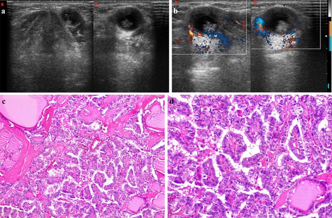
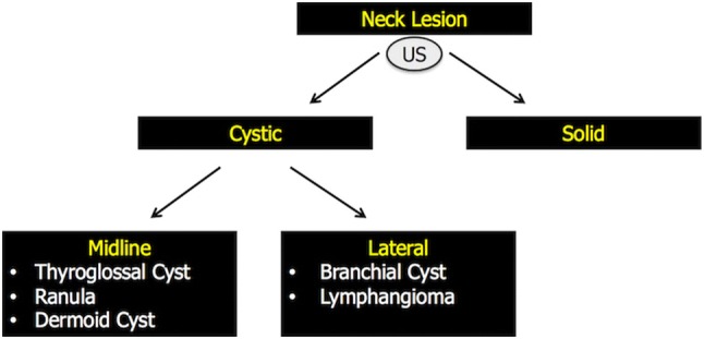
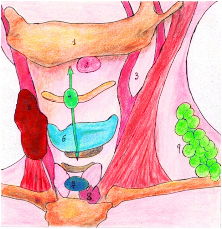
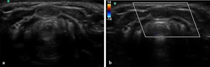
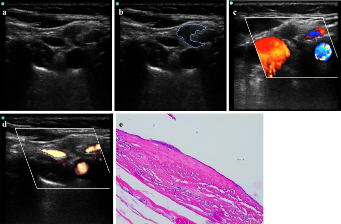
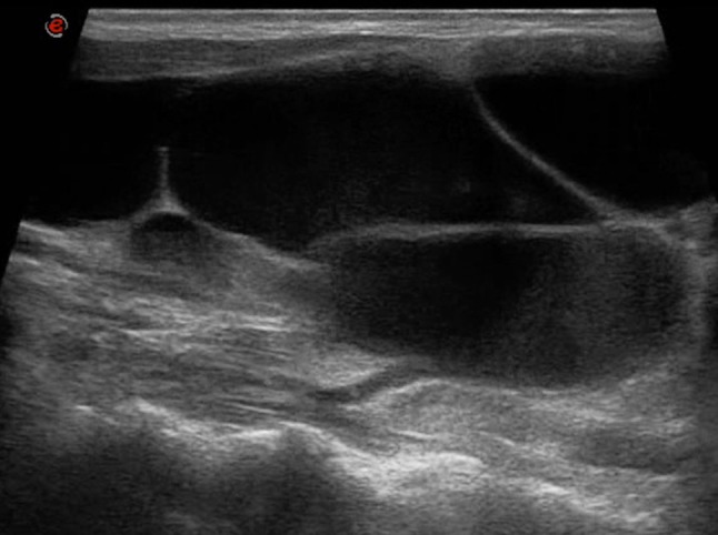
Thyroglossal duct cysts (TGDCs) are the most common congenital abnormality of the neck, accounting for approximately 70% of congenital neck lesions. Two-thirds of thyroglossal duct anomalies are diagnosed within the first three decades of life, with more than half being identified before 10 years of age. The age of presentation, clinical examination and imaging are essential for an accurate diagnosis. This review aims to summarize the imaging findings of TGDCs and their main differential diagnoses with emphasis on ultrasound assessment. A focus on site-specific key differentiating between them is also addressed.

Keywords: Cystic neck lesions; Doppler techniques; Magnetic resonance imaging; Neck imaging; Neck ultrasound.


Conflict of interest statement

The authors have no conflicts of interest.

Figures

Fig. 1
Fig. 1
Schematic diagram illustrating the anatomic pathway of TGD. It originates from the foramen caecum (F) of the tongue, descends in the midline first anteriorly and then looping superiorly behind the hyoid bone (H). It descends further downwards anterior to the thyrohyoid membrane, thyroid cartilage (TC) and reaches its final position at the thyroid gland (TG)
Fig. 2
Fig. 2
TGDC in a 44-year-old male with a mass in the midline of neck and erythema of the overlying skin. B-mode US (a) and PD (b) images show a homogeneous hypoechoic lesion with diffuse low-level internal echoes. Increased through transmission is present. No internal flow is observed with PD. Pathologic examination of surgical specimen showed a TGDC with infection-inflammation, lined by squamous epithelium
Fig. 3
Fig. 3
Complicated TGDC in a 21-year-old boy, slightly off-midline to the left, between the thyroid cartilage and strap muscles. Transverse B-mode US image (on the left) show a uniformly echogenic, pseudosolid appearance of a TGDC. Note, however, the posterior enhancement, suggesting its cystic nature. No internal flow is observed with CD (in the middle). SE (on the right) shows elasticity in the whole region of interest (homogeneously green with some red areas mixed in), an appearance suggestive of benign lesion. TGDC was confirmed by pathologic examination of surgical specimen, which showed no infection or hemorrhage. Pseudosolid pattern was due to proteinaceous content of cyst
Fig. 4
Fig. 4
Young male (19-year-old) with a midline neck swelling, moving with deglutition. B-mode US (a) and PD (b) images show a well-circumscribed, heterogenous complex cystic lesion containing a solid-appearing echogenic nodularity due to a previous hemorrhage in the midline of neck, at the level of hyoid bone. Increased through transmission is present. No internal flow is observed with PD. A MRI of the neck was subsequently done, which demonstrated a large midline TGDC, attached to the body of the hyoid bone near the foramen cecum of the tongue base. Neck axial T1—(c) and T2-weighted fat-suppressed (d) images at the level of hyod bone and midline sagittal T2-weighted fat-suppressed (e) images show the high signal TGDC in a prevalent infrahyoid location with a superior component extending directly posterior to the hyoid bone. Axial T1—(f) and T2-weighted (g) images between the level of hyoid bone and thyroid cartilage allow to demonstrate the TGDC embedded in the left-sided strap muscles, a characteristic cross-sectional finding of TGDCs. h, i Photomicrographs—hematoxylin–eosin stain, magnification × 4 (h) and  × 20 (i). TGDC characterized by pseudostratified ciliated epithelium and inflammatory stromal infiltrate; cystic content was found to be haemorragic
Fig. 5
Fig. 5
Infrahyoid TDGC with malignancy in a 34-year-old man, slightly off-midline to the left, between the thyroid cartilage and strap muscles. B-Mode US (a) and color-Doppler (b) images reveal a well-circumscribed anechoic structure with a grossly irregular nodular component in its inferior portion, which shows vascular signals on Doppler imaging. Posterior acoustic enhancement is also evident. Resection revealed a papillary carcinoma(c,  × 10 and d,  × 20)
Fig. 6
Fig. 6
Evidence-based diagnostic algorithm proposed to guide clinicians and radiologists in the correct differential diagnosis of neck cystic lesions [40]. Briefly, if a neck lesion is detected, US can usually determine whether it is cystic or solid. Then, once established that the lesion is cystic, its location will often point to its nature. If located on the midline, the differential diagnosis narrows to TGDCs, ranulas or dermoid cysts. Off-midline lesions can be, instead, branchial cleft cysts or lymphangiomas
Fig. 7
Fig. 7
Midline lesions are either thyroglossal duct cysts, dermoid cysts or ranulas. Off-midline lesions can be branchial cleft cysts or lymphangiomas. (1) Mandible; (2) Ranula; (3) Sternocleidomastoid muscle—anterior border, lateral to the carotid artery bifurcation, space of lateral neck most frequently occupied by branchial cysts; (4)TGDC; (5)Fibromatosis colli; (6)Thyroid cartilage; (7) Dermoid cyst; (8) Thymus; (9) Lymphangioma
Fig. 8
Fig. 8
Dermoid cyst in a 38-year-old woman. a Transverse B-mode US image shows a well-defined, unilocular, homogeneously hypoechoic lesion with internal echogenic dots, midline in location and superficial to the strap muscles (thyrohyoid muscles) at the cross-section level of cricoid cartilage. b Corresponding CD images show no intenal flow
Fig. 9
Fig. 9
Second BCC in a 42-year-old girl, incidentally discovered during a screening US examination for thyroid pathologies. B-mode US (a) and corresponding US schematic (b) images of the lateral neck shows a small, well-marginated, anechoic lesion with thin walls and internal fine echoes along the anterior border of the sternocleidomastoid muscle, lateral to the carotid bifurcation. A “beak sign” is seen as a curved rim of the lesion pointing medially. c, d CD e PD landmarks (carotid bifurcation, giugular vein) are useful to better identify the BCC. e Photomicrograph–hematoxylin–eosin stain, magnification × 4. Second BCC lined by squamous epithelium
Fig. 10
Fig. 10
B-mode US image shows a large cystic lesion with internal septa and debris centered in the left neck base compatible with a MLM

References

    1. Patel S, Bhatt AA. Thyroglossal duct pathology and mimics. Insights Imaging. 2019;10(1):12. doi: 10.1186/s13244-019-0694-x. - DOI - PMC - PubMed
    1. Zander DA, Smoker WR. Imaging of ectopic thyroid tissue and thyroglossal duct cysts. Radiographics. 2014;34(1):37–50. doi: 10.1148/rg.341135055. - DOI - PubMed
    1. Wadsworth DT, Siegel MJ. Thyroglossal duct cysts: variability of sonographic findings. AJR Am J Roentgenol. 1994;163(6):1475–1477. doi: 10.2214/ajr.163.6.7992750. - DOI - PubMed
    1. Dedivitis RA, Camargo DL, Peixoto GL, Weissman L, Guimarães AV. Thyroglossal duct: a review of 55 cases. J Am Coll Surg. 2002;194(3):274–277. doi: 10.1016/S1072-7515(01)01171-1. - DOI - PubMed
    1. Brousseau VJ, Solares CA, Xu M, Krakovitz P, Koltai PJ. Thyroglossal duct cysts: presentation and management in children versus adults. Int J Pediatr Otorhinolaryngol. 2003;67(12):1285–1290. doi: 10.1016/j.ijporl.2003.07.006. - DOI - PubMed

LinkOut - more resources