

The genetic and clinical landscape of nanophthalmos and posterior microphthalmos in an Australian cohort
- PMID: 32052405
- PMCID: PMC7811993
- DOI: 10.1111/cge.13722
The genetic and clinical landscape of nanophthalmos and posterior microphthalmos in an Australian cohort
Abstract
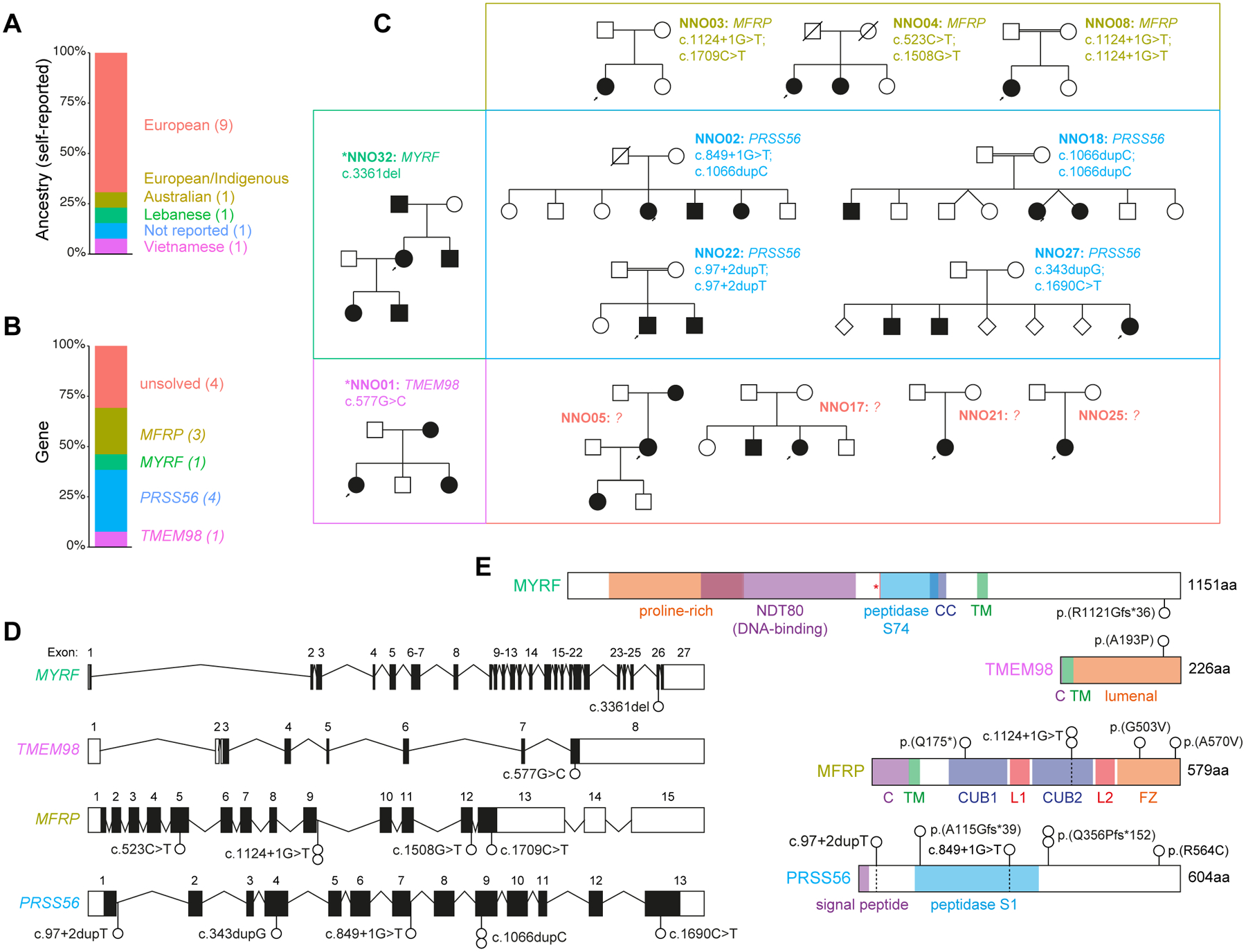
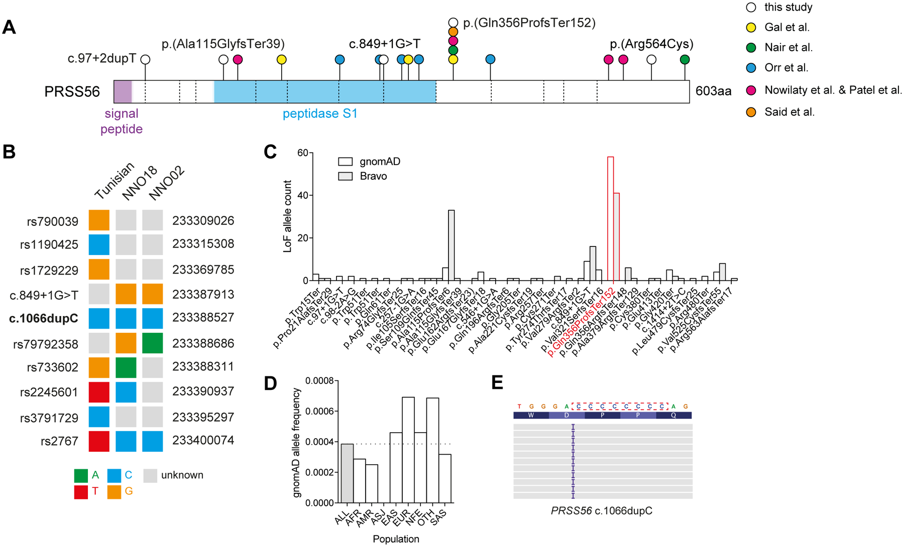
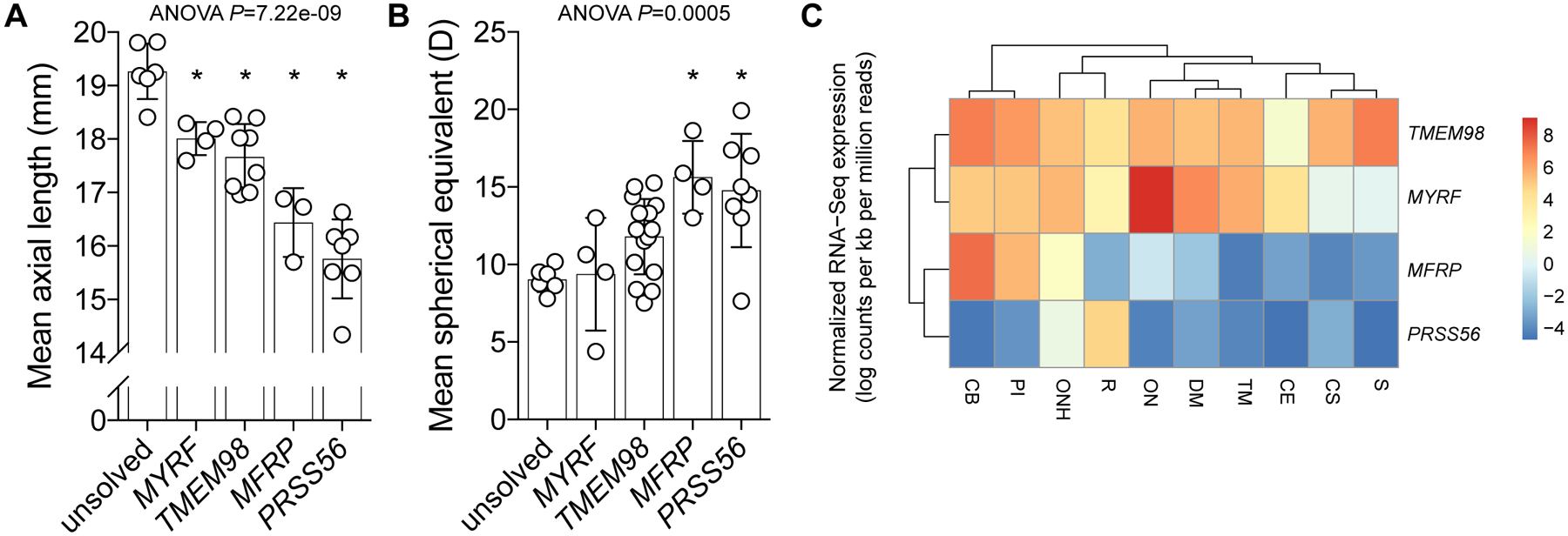
Nanophthalmos and posterior microphthalmos are ocular abnormalities in which both eyes are abnormally small, and typically associated with extreme hyperopia. We recruited 40 individuals from 13 kindreds with nanophthalmos or posterior microphthalmos, with 12 probands subjected to exome sequencing. Nine probands (69.2%) were assigned a genetic diagnosis, with variants in MYRF, TMEM98, MFRP, and PRSS56. Two of four PRSS56 families harbored the previously described c.1066dupC variant implicated in over half of all reported PRSS56 kindreds, with different surrounding haplotypes in each family suggesting a mutational hotspot. Individuals with a genetic diagnosis had shorter mean axial lengths and higher hyperopia than those without, with recessive forms associated with the most extreme phenotypes. These findings detail the genetic architecture of nanophthalmos and posterior microphthalmos in a cohort of predominantly European ancestry, their relative clinical phenotypes, and highlight the shared genetic architecture of rare and common disorders of refractive error.
Keywords: MFRP; MYRF; PRSS56; TMEM98; axial length; microphthalmia; nanophthalmos; posterior microphthalmos.
© 2020 John Wiley & Sons A/S. Published by John Wiley & Sons Ltd.
Conflict of interest statement
Figures
Similar articles
-
Novel TMEM98, MFRP, PRSS56 variants in a large United States high hyperopia and nanophthalmos cohort.Sci Rep. 2020 Nov 17;10(1):19986. doi: 10.1038/s41598-020-76725-8. Sci Rep. 2020. PMID: 33203948 Free PMC article.
-
Autosomal dominant nanophthalmos and high hyperopia associated with a C-terminal frameshift variant in MYRF.Mol Vis. 2019 Sep 21;25:527-534. eCollection 2019. Mol Vis. 2019. PMID: 31700225 Free PMC article.
-
Novel TMEM98 mutations in pedigrees with autosomal dominant nanophthalmos.Mol Vis. 2015 Sep 1;21:1017-23. eCollection 2015. Mol Vis. 2015. PMID: 26392740 Free PMC article.
-
Glaucoma Syndromes: Insights into Glaucoma Genetics and Pathogenesis from Monogenic Syndromic Disorders.Genes (Basel). 2021 Sep 11;12(9):1403. doi: 10.3390/genes12091403. Genes (Basel). 2021. PMID: 34573386 Free PMC article. Review.
-
MFRP variant results in nanophthalmos, retinitis pigmentosa, variability in foveal avascular zone.Ophthalmic Genet. 2023 Feb;44(1):83-88. doi: 10.1080/13816810.2022.2103835. Epub 2022 Jul 26. Ophthalmic Genet. 2023. PMID: 35880649 Review.
Cited by
-
Insight into small eyes: a practical description from phenotypes presentations to the management.Int J Ophthalmol. 2024 Feb 18;17(2):380-391. doi: 10.18240/ijo.2024.02.23. eCollection 2024. Int J Ophthalmol. 2024. PMID: 38371260 Free PMC article. Review.
-
MFRP variations cause nanophthalmos in five Chinese families with distinct phenotypic diversity.Front Genet. 2024 Jul 15;15:1407361. doi: 10.3389/fgene.2024.1407361. eCollection 2024. Front Genet. 2024. PMID: 39076172 Free PMC article.
-
A Novel Mutation in the Membrane Frizzled-Related Protein Gene for Posterior Microphthalmia, Non-pigmented Retinitis Pigmentosa, Optic Nerve Drusen, and Retinoschisis in a Consanguineous Family.Front Med (Lausanne). 2022 Mar 24;9:835621. doi: 10.3389/fmed.2022.835621. eCollection 2022. Front Med (Lausanne). 2022. PMID: 35402469 Free PMC article.
-
The Pathogenesis and Treatment of Complications in Nanophthalmos.J Ophthalmol. 2020 Jul 19;2020:6578750. doi: 10.1155/2020/6578750. eCollection 2020. J Ophthalmol. 2020. PMID: 32765903 Free PMC article. Review.
-
Genetic Spectrum and Genotype-Phenotype Correlations in a Chinese Cohort With Nanophthalmos With Secondary Angle-Closure Glaucoma.Invest Ophthalmol Vis Sci. 2025 Jun 2;66(6):9. doi: 10.1167/iovs.66.6.9. Invest Ophthalmol Vis Sci. 2025. PMID: 40459495 Free PMC article.
References
-
- Bourne RRA, Stevens GA, White RA, et al. Causes of vision loss worldwide, 1990–2010: a systematic analysis. Lancet Glob Health. 2013;1(6):e339–e349. - PubMed
-
- Khan AO. Posterior microphthalmos versus nanophthalmos. Ophthalmic Genet. 2008;29(4):189. - PubMed
-
- Aldahmesh MA, Nowilaty SR, Alzahrani F, et al. Posterior microphthalmos as a genetically heterogeneous condition that can be allelic to nanophthalmos. Arch Ophthalmol. 2011;129(6):805–807. - PubMed
-
- Nowilaty SR, Khan AO, Aldahmesh MA, Tabbara KF, Al-Amri A, Alkuraya FS. Biometric and molecular characterization of clinically diagnosed posterior microphthalmos. Am J Ophthalmol. 2013;155(2):361–372.e7. - PubMed
Publication types
MeSH terms
Substances
Supplementary concepts
Grants and funding
LinkOut - more resources
Full Text Sources
Miscellaneous
