Review
. 2020 Apr;28(2):79-93.
doi: 10.4250/jcvi.2019.0103. Epub 2020 Jan 13.

Myocardial Positron Emission Tomography for Evaluation of Cardiac Sarcoidosis: Specialized Protocols for Better Diagnosis

Affiliations
Review

Myocardial Positron Emission Tomography for Evaluation of Cardiac Sarcoidosis: Specialized Protocols for Better Diagnosis

In Chang Hwang et al. J Cardiovasc Imaging. 2020 Apr.

Abstract

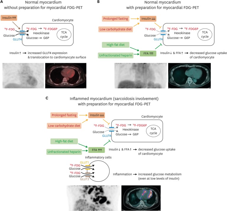
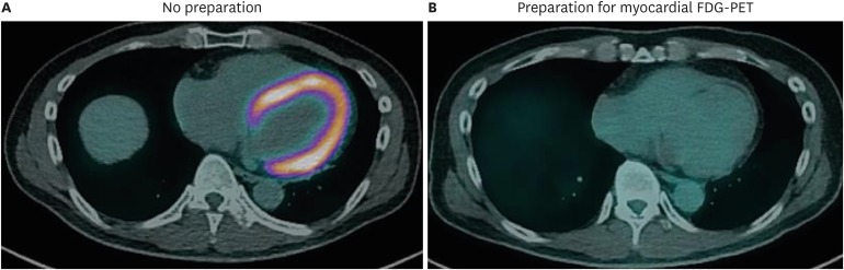
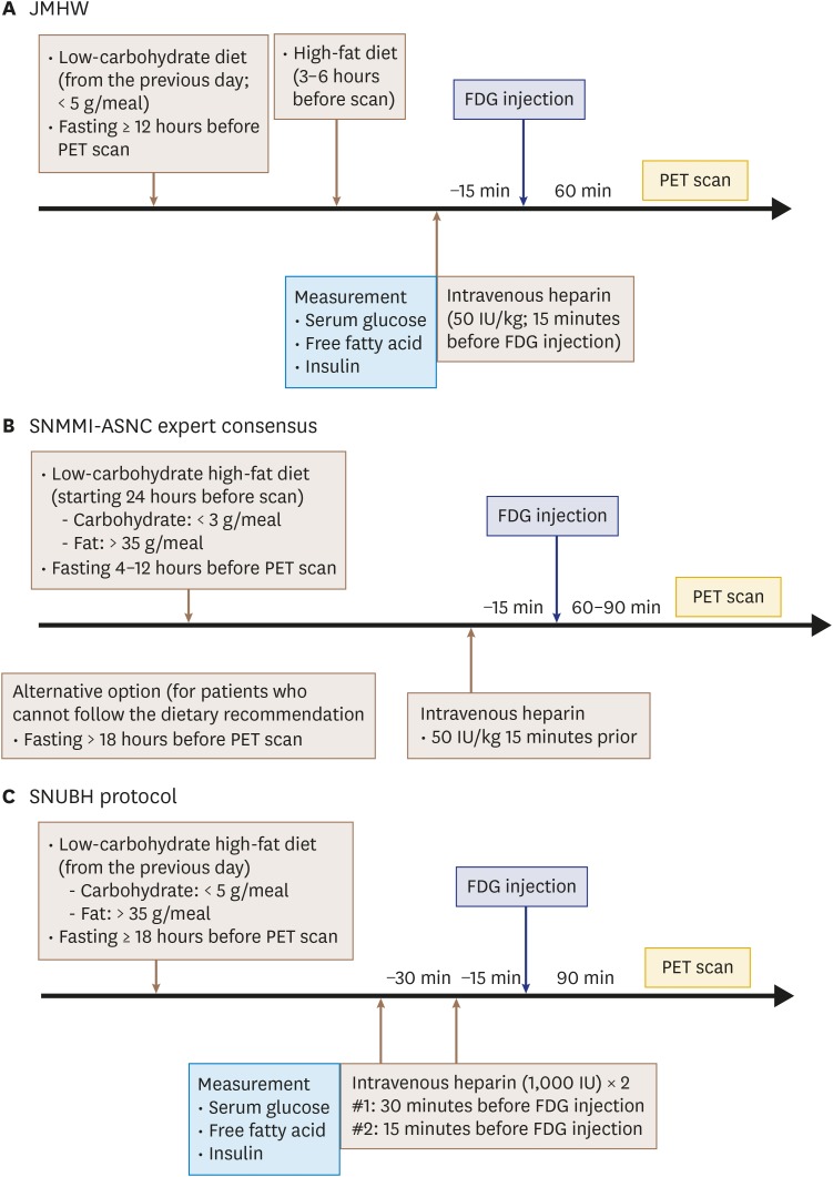
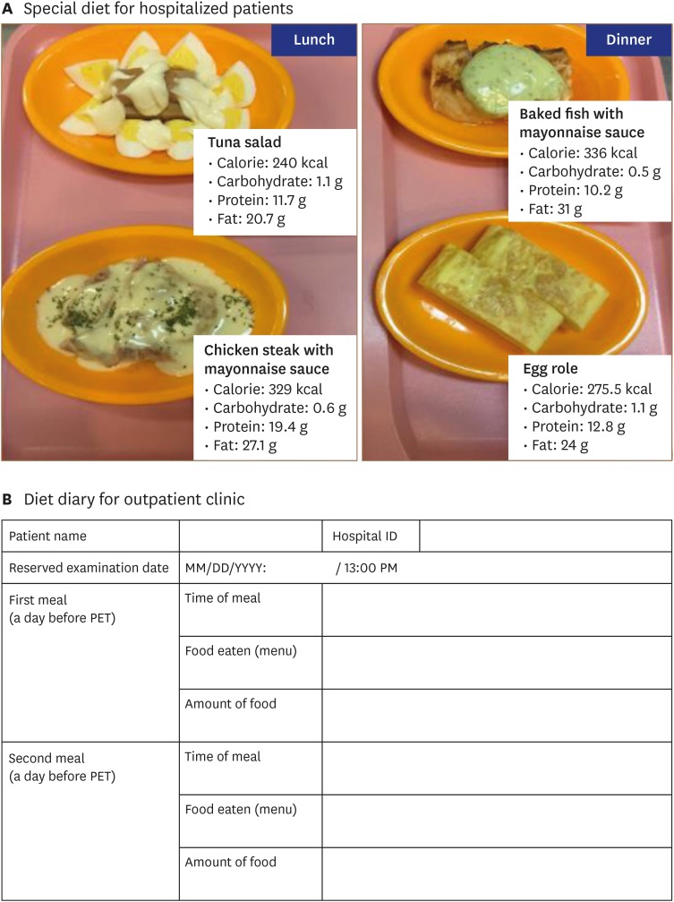
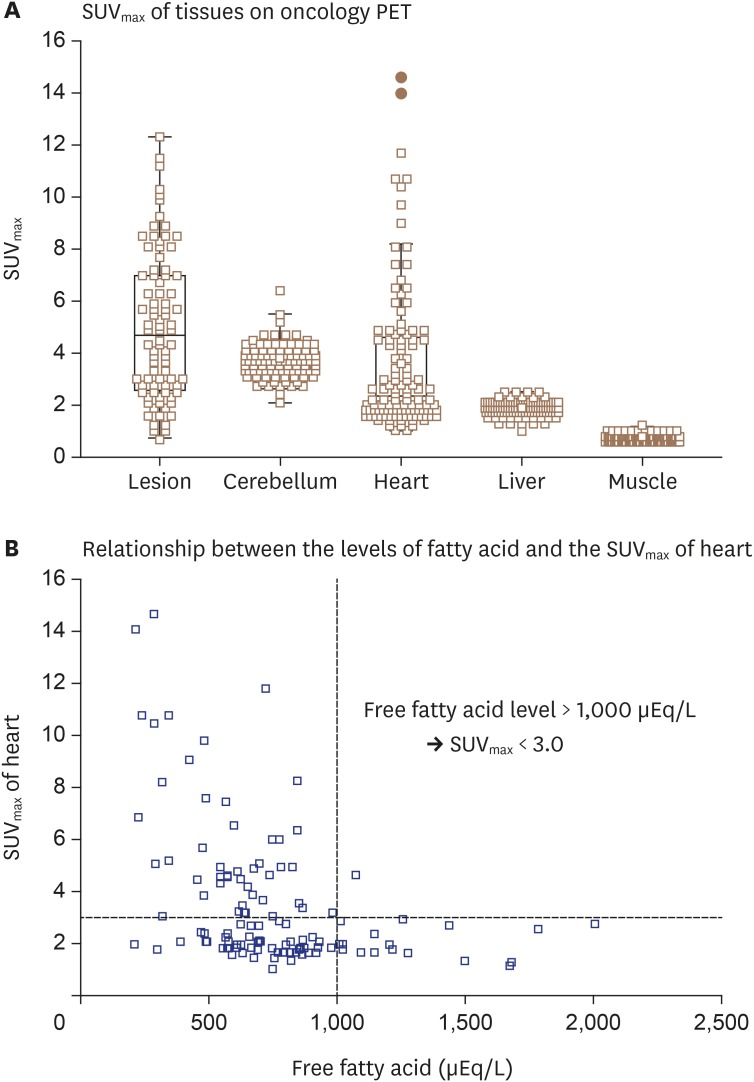
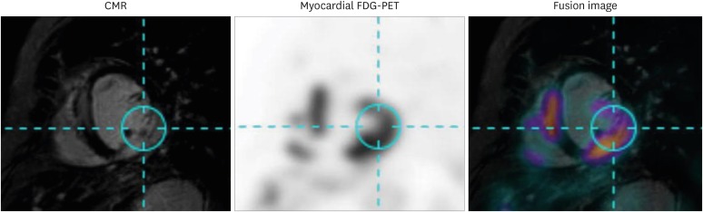
Sarcoidosis is a multisystemic granulomatous disease of unknown etiology with various clinical presentations depending on the organs involved. Since cardiac sarcoidosis (CS) portends a higher risk of morbidity and mortality, early diagnosis and aggressive medical treatment are essential to improve the prognosis. 18F-Fluorodeoxyglucose (FDG) positron emission tomography (PET) has emerged as an important tool with practical advantages in assessing disease activity and monitoring the treatment response in patients with CS. While it has high sensitivity, it also has great variability in specificity, probably due to normal physiologic myocardial FDG uptake, which interferes with the evaluation and follow-up of CS using FDG-PET. This review details the technical aspects of FDG-PET imaging for evaluating and diagnosing CS, assessing disease activity, and monitoring therapeutic response.

Keywords: Cardiac sarcoidosis; Extended fasting; High-fat low-carbohydrate diet; Multimodality imaging; Myocardial positron emission tomography.


Conflict of interest statement

The authors have no financial conflicts of interest.

Figures

Figure 1
Figure 1. Physiologic myocardium FDG uptake and detection of inflammatory lesions on FDG-PET. Schematic figures detecting physiologic glucose uptake in normal myocardium (A) and suppression of glucose uptake by dietary preparations, prolonged fasting, and unfractionated heparin administration (B). (C) Appropriate preparation for myocardial FDG-PET can suppress physiologic glucose uptake in cardiomyocytes while maintaining glucose uptake of inflammatory cells in the myocardium with sarcoidosis involvement. FDG: fluorodeoxyglucose, FDG6P: fluorodeoxyglucose-6-phosphate, FFA: free fatty acid, G6P: glucose-6-phosphate, GLUT: glucose transporter, PET: positron emission tomography, TCA: tricarboxylic acid.
Figure 2
Figure 2. Suppression of physiologic glucose uptake in normal myocardium. FDG-PET/CT images of patient with suspected cardiac sarcoidosis. (A) This patient underwent initial PET/CT scan with 18 hours of fasting, without a dietary preparation, and showed diffusely increased metabolism in the whole myocardium. (B) During the same hospitalization, this patient underwent PET/CT scanning again after a low-carbohydrate high-fat diet and 18 hours of fasting. PET/CT image after appropriate preparation showed no abnormal uptake in the myocardium, as it was successfully suppressed by the dietary preparation. CT: computed tomography, FDG: fluorodeoxyglucose: PET: positron emission tomography.
Figure 3
Figure 3. Suggested protocols for diagnosing cardiac sarcoidosis. Suggested myocardial FDG-PET protocols for diagnosing cardiac sarcoidosis provided by the Japanese Ministry of Health and Welfare (JMHW) (A), and the Joint Society of Nuclear Medicine and Molecular Imaging (SNMMI) and American Society of Nuclear Cardiology (ASNC) expert consensus statement (B) are summarized. (C) Protocol used by Seoul National University Bundang Hospital (SNUBH) is also shown. FDG: fluorodeoxyglucose, PET: positron emission tomography.
Figure 4
Figure 4. Dietary preparations for patients with suspected cardiac sarcoidosis undergoing myocardial PET. (A) Special diets for hospitalized patients with suspected or known cardiac sarcoidosis undergoing myocardial PET. The day before FDG-PET scan, patients are provided lunch (left) and dinner (right) that are low in carbohydrates (< 5 g/meal) and high in fat (> 35 g/meal), as developed by physicians and a nutritional support team. After consuming dinner at 18:00 PM, patients fast for 18 hours, and PET scan is performed at 13:00 PM. (B) Sample diet diary for patient at out-patient clinic. PET: positron emission tomography.
Figure 5
Figure 5. Myocardial FDG uptake among 147 patients who underwent oncology PET. Serum free fatty acid level and heart SUVmax were measured in 147 patients who underwent FDG-PET for diagnosis of malignancy. (A) On FDG-PET without preparation for sarcoidosis evaluation, the heart and brain demonstrate higher SUVmax values than liver or muscle. (B) Serum free fatty acid level at time of FDG-PET shows inverse correlation with heart SUVmax, suggesting that serum fatty acid level > 1,000 μEq/L is needed at the time of myocardial FDG-PET imaging to suppress physiologic uptake of glucose in normal myocardium. FDG: fluorodeoxyglucose, PET: positron emission tomography, SUVmax: maximal standardized uptake value.
Figure 6
Figure 6. Representative case of hybrid CMR-PET imaging. CMR and myocardial FDG-PET images of patient with cardiac sarcoidosis are shown. Images of these two modalities were fused using dedicated software and show co-localization of late gadolinium enhancement on CMR and increased FDG uptake on myocardial FDG-PET. CMR: cardiac magnetic resonance, FDG: fluorodeoxyglucose, PET: positron emission tomography.

References

    1. Porter GH. Sarcoid heart disease. N Engl J Med. 1960;263:1350–1357. - PubMed
    1. Birnie DH, Nery PB, Ha AC, Beanlands RS. Cardiac Sarcoidosis. J Am Coll Cardiol. 2016;68:411–421. - PubMed
    1. Silverman KJ, Hutchins GM, Bulkley BH. Cardiac sarcoid: a clinicopathologic study of 84 unselected patients with systemic sarcoidosis. Circulation. 1978;58:1204–1211. - PubMed
    1. Bennett MK, Gilotra NA, Harrington C, et al. Evaluation of the role of endomyocardial biopsy in 851 patients with unexplained heart failure from 2000-2009. Circ Heart Fail. 2013;6:676–684. - PubMed
    1. Birnie DH, Sauer WH, Bogun F, et al. HRS expert consensus statement on the diagnosis and management of arrhythmias associated with cardiac sarcoidosis. Heart Rhythm. 2014;11:1305–1323. - PubMed