. 2020 Mar;19(5):610-624.
doi: 10.1080/15384101.2020.1728024. Epub 2020 Feb 13.

Long noncoding RNA LINC00963 promotes breast cancer progression by functioning as a molecular sponge for microRNA-625 and thereby upregulating HMGA1

Affiliations

Long noncoding RNA LINC00963 promotes breast cancer progression by functioning as a molecular sponge for microRNA-625 and thereby upregulating HMGA1

Zhen Wu et al. Cell Cycle. 2020 Mar.

Abstract

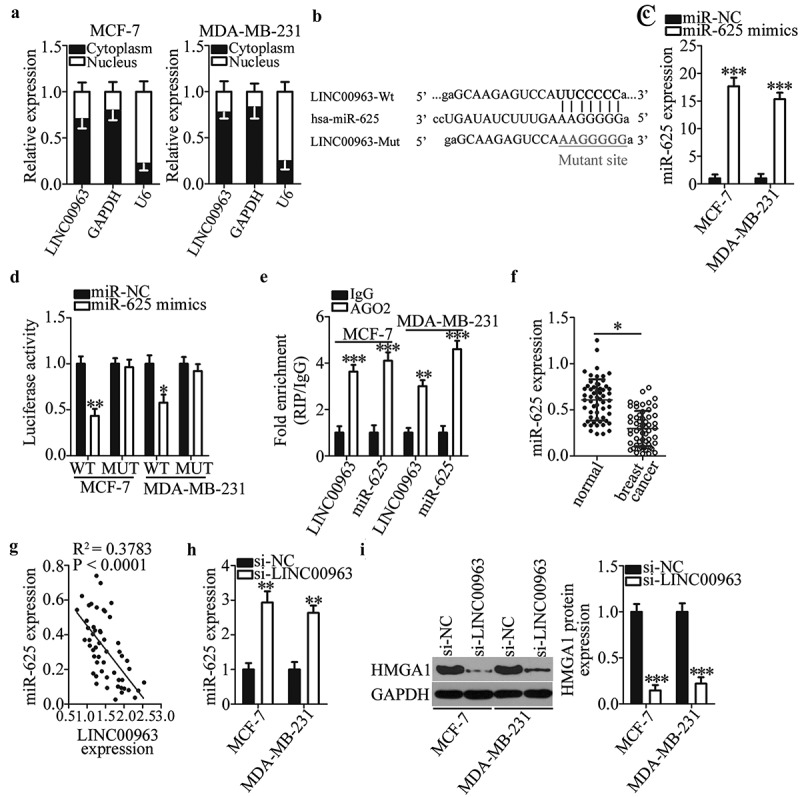
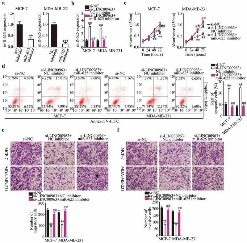
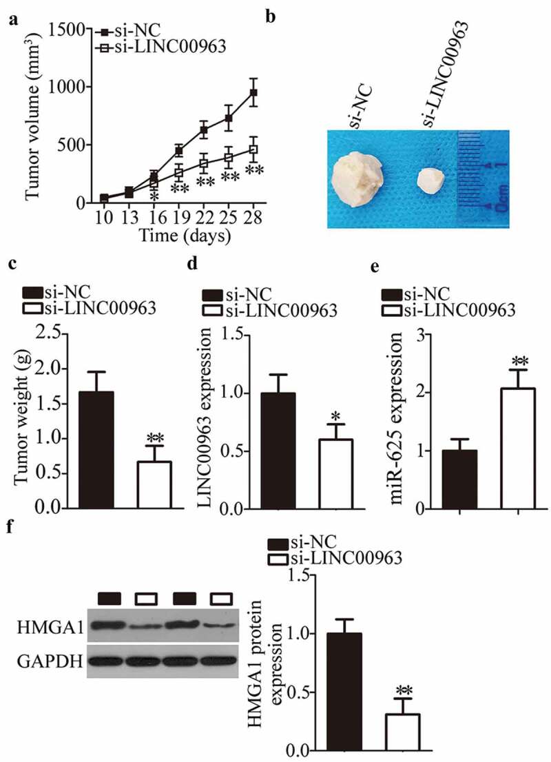
Extensive research has shown that LINC00963 is aberrantly expressed in human cancers, and that dysregulation of LINC00963 is implicated in the initiation and progression of human cancers. The expression and functions of LINC00963 in breast cancer are still unclear. Our aims were to measure the expression of LINC00963 in breast cancer, determine its effects on malignant behaviors of tumor cells, and uncover the molecular events underlying the actions of LINC00963 in breast cancer. Herein, LINC00963 was found to be overexpressed in breast cancer samples, and its overexpression was correlated with lymph node metastasis, TNM stage and differentiation grade. Patients with breast cancer harboring higher LINC00963 expression showed shorter overall survival than did the patients with lower LINC00963 expression. Functional experiments revealed that depletion of LINC00963 inhibited breast cancer cell proliferation, migration, and invasion and facilitated apoptosis in vitro and impaired tumor growth in vivo. Mechanism investigation revealed that LINC00963 can interact with microRNA-625 (miR-625). LINC00963 worked as a competitive endogenous RNA for miR-625 to weaken the suppressive effect of miR-625 on high mobility group AT-hook 1 (HMGA1) in breast cancer cells. Furthermore, miR-625 inhibition and HMGA1 restoration both abrogated the effects of LINC00963 silencing on breast cancer cells. Our findings indicate that the LINC00963-miR-625-HMGA1 pathway plays an important role in the malignancy of breast cancer in vitro and in vivo. Hence, targeting this pathway may be a novel strategy against breast cancer.

Keywords: High mobility group AT-hook 1; LINC00963; breast cancer; microRNA-625.


Figures

Figure 1.
Figure 1.
LINC00963 expression is excessive in breast cancer and predicts poorer prognosis. (a) RT-qPCR analysis was performed to determine LINC00963 expression in 53 pairs of breast cancer tissue samples and matched adjacent normal tissues. Paired student’s t-test is utilized for statistical analysis. **P < 0.01 vs. adjacent normal tissues. (b) Expression of LINC00963 in four breast cancer cell lines (BT-474, MCF-7, MDA-MB-231, and SKBR3) and in human normal breast epithelial cell line MCF-10A was tested via RT-qPCR. Unpaired student’s t-test is utilized for statistical analysis. *P < 0.05, **P < 0.01 vs. MCF-10A. (c) The Kaplan–Meier method and logrank test were utilized to analyze the correlation between LINC00963 expression and the overall survival among patients with breast cancer. The overall-survival curve was analyzed by the Kaplan–Meier method, and the log-rank test was conducted to compare the survival data. P = 0.040.
Figure 2.
Figure 2.
LINC00963 knockdown attenuates proliferation, migration, and invasiveness and increases apoptosis of breast cancer cells. (a) Si-LINC00963 was designed to successfully knock down endogenous LINC00963 in MCF-7 and MDA-MB-231 cells. ***P < 0.001 vs. si-NC. (b) The CCK-8 assay shows the impact of the LINC00963 knockdown on the proliferation of MCF-7 and MDA-MB-231 cells. **P < 0.05 vs. si-NC. (c) Investigation of the apoptosis rate of LINC00963-depleted MCF-7 and MDA-MB-231 cells was performed by flow cytometry. **P < 0.05 vs. si-NC. (d, e) Influences of LINC00963 silencing on the migratory and invasive abilities of MCF-7 and MDA-MB-231 cells were evaluated by Transwell migration and invasion assays. **P < 0.01 vs. si-NC. Unpaired student’s t-test is utilized for statistical analysis.
Figure 3.
Figure 3.
LINC00963 functions as a sponge for miR-625 in breast cancer cells. (a) LINC00963 is mainly expressed in the cytoplasm of MCF-7 and MDA-MB-231 cells. (b) The wild-type miR-625–binding sequence in LINC00963 was predicted by starBase 3.0. The mutant miR-625–binding sequences are shown too. (c) The miR-625 mimics was introduced into MCF-7 and MDA-MB-231 cells to elevate the endogenous miR-625 expression. Unpaired student’s t-test is utilized for statistical analysis. ***P < 0.001 vs. miR-NC. (d) The luciferase reporter assay was conducted to confirm the interaction between miR-625 and LINC00963 in breast cancer cells. Luciferase activities were measured in MCF-7 and MDA-MB-231 cells after cotransfection with either the miR-625 mimics or miR-NC and either LINC00963-WT or LINC00963-MUT. Unpaired student’s t-test is utilized for statistical analysis. *P < 0.05, **P < 0.01 vs. miR-NC. (e) The RIP assay indicates that LINC00963 and miR-625 are preferentially enriched by the anti-AGO2 antibody after immunoprecipitation in the lysates of MCF-7 and MDA-MB-231 cells. Unpaired student’s t-test is utilized for statistical analysis. **P < 0.01, ***P < 0.001 vs. IgG. (f) MiR-625 expression in 53 pairs of breast cancer tissue samples and matched adjacent normal tissue samples, as determined by RT-qPCR. *P < 0.05 vs. adjacent normal tissues. Paired student’s t-test is utilized for statistical analysis. (g) Spearman’s correlation analysis was carried out to test the expression correlations between LINC00963 and miR-625 in breast cancer tissues. R2 = 0.3783, P < 0.0001. (h) The impact of the LINC00963 knockdown on miR-625 expression was examined in MCF-7 and MDA-MB-231 cells after either si-LINC00963 or si-NC transfection. Unpaired student’s t-test is utilized for statistical analysis. **P < 0.01 vs.si-NC. (i) Western blotting revealed that HMGA1 protein expression is suppressed by si-LINC00963 in MCF-7 and MDA-MB-231 cells. Unpaired student’s t-test is utilized for statistical analysis. ***P < 0.001 vs. si-NC.
Figure 4.
Figure 4.
A reduction in LINC00963 expression suppresses the proliferation, migration, and invasiveness and accelerates the apoptosis of breast cancer cells via direct modulation of miR-625. (a) MCF-7 and MDA-MB-231 cells transfected with either the miR-625 inhibitor or NC inhibitor were collected and subjected to RT-qPCR analysis for the determination of miR-625 expression. Unpaired student’s t-test is utilized for statistical analysis. **P < 0.01, ***P < 0.001 vs. NC inhibitor. (b) Si-LINC00963 and either the miR-625 inhibitor or NC inhibitor were cotransfected into MCF-7 and MDA-MB-231 cells. After 48 h culture, RT-qPCR analysis was conducted to assess miR-625 expression. *P < 0.01 vs. si-NC. ##P < 0.01vs. si-LINC00963+ NC inhibitor. (c, d) The CCK-8 assay and flow-cytometric analysis were performed to prove that the effects of si-LINC00963 on the proliferation and apoptosis of MCF-7 and MDA-MB-231 cells are abrogated by the miR-625 inhibitor. **P < 0.01 vs. si-NC. ##P < 0.01 vs. si-LINC00963+ NC inhibitor. (e, f) Transwell migration and invasion assays were conducted to analyze the migration and invasion status of MCF-7 and MDA-MB-231 cells cotransfected with si-LINC00963 and either the miR-625 inhibitor or NC inhibitor. **P < 0.01 vs. si-NC. ##P < 0.05 vs. si-LINC00963+ NC inhibitor. One-way analysis of variance followed by Tukey’s post hoc test was utilized for statistical analysis.
Figure 5.
Figure 5.
Recovery of HMGA1 expression rescinds the effects of the LINC00963 knockdown on MCF-7 and MDA-MB-231 cells. Either the HMGA1-overexpressing plasmid (pc-HMGA1) or the empty pcDNA3.1 vector in combination with si-LINC00963 was introduced into MCF-7 and MDA-MB-231 cells. The transfected cells were studied in the following experiments. (a) The protein expression of HMGA1 in MCF-7 and MDA-MB-231 cells treated as described above was examined by western blotting. ***P < 0.001 vs. si-NC. ###P < 0.001 vs. si-LINC00963+ pcDNA3.1. (b–e) The CCK-8 assay, flow cytometry, and Transwell migration and invasion assays were carried out to determine the proliferation, apoptosis, migration, and invasiveness of MCF-7 and MDA-MB-231 cells that were cotransfected with si-LINC00963 and either pc-HMGA1 or pcDNA3.1. **P < 0.01 vs. si-NC. ##P < 0.01 vs. si-LINC00963+ pcDNA3.1. One-way analysis of variance followed by Tukey’s post hoc test was utilized for statistical analysis.
Figure 6.
Figure 6.
LINC00963 silencing restrains breast tumor growth in vivo. (a) MCF-7 cells transfected with either si-LINC00963 or si-NC were injected into nude mice. Tumor growth curves revealed that the tumor xenografts grew at a markedly slower rate in the si-LINC00963 group than in the si-NC group. *P < 0.05. **P < 0.01 vs. si-NC. (b) At the end of this experiment, the mice were anesthetized, and tumor xenografts were excised. The representative images of tumor xenografts are presented. (c) The weight of tumor xenografts obtained from groups si-LINC00963 and si-NC was measured. **P < 0.01 vs. si-NC. (d, e) The levels of LINC00963 and miR-625 in the tumor xenografts were assessed by RT-qPCR. *P < 0.05, **P < 0.01 vs. si-NC. (f) Western blotting was carried out to measure HMGA1 protein expression in the tumor xenografts. **P < 0.01 vs. si-NC. Unpaired student’s t-test is used for all above statistical analysis.

References

    1. Siegel RL, Miller KD, Jemal A.. Cancer statistics, 2018. CA Cancer J Clin. 2018;68:7–30. - PubMed
    1. Fan L, Strasser-Weippl K, Li JJ, et al. Breast cancer in China. Lancet Oncol. 2014;15:e279–89. - PubMed
    1. Liu H, Ye H.. Screening of the prognostic targets for breast cancer based co-expression modules analysis. Mol Med Rep. 2017;16:4038–4044. - PMC - PubMed
    1. Cancer Genome Atlas N . Comprehensive molecular portraits of human breast tumours. Nature. 2012;490:61–70. - PMC - PubMed
    1. Lehmann BD, Bauer JA, Chen X, et al. Identification of human triple-negative breast cancer subtypes and preclinical models for selection of targeted therapies. J Clin Invest. 2011;121:2750–2767. - PMC - PubMed

MeSH terms