. 2020 May;160(1):52-63.
doi: 10.1111/imm.13180. Epub 2020 Mar 17.

Expression of melatonin receptors and CD4 in the ovine thymus, lymph node, spleen and liver during early pregnancy

Affiliations

Expression of melatonin receptors and CD4 in the ovine thymus, lymph node, spleen and liver during early pregnancy

Jiachen Bai et al. Immunology. 2020 May.

Abstract

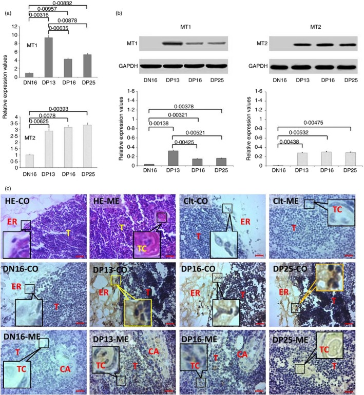
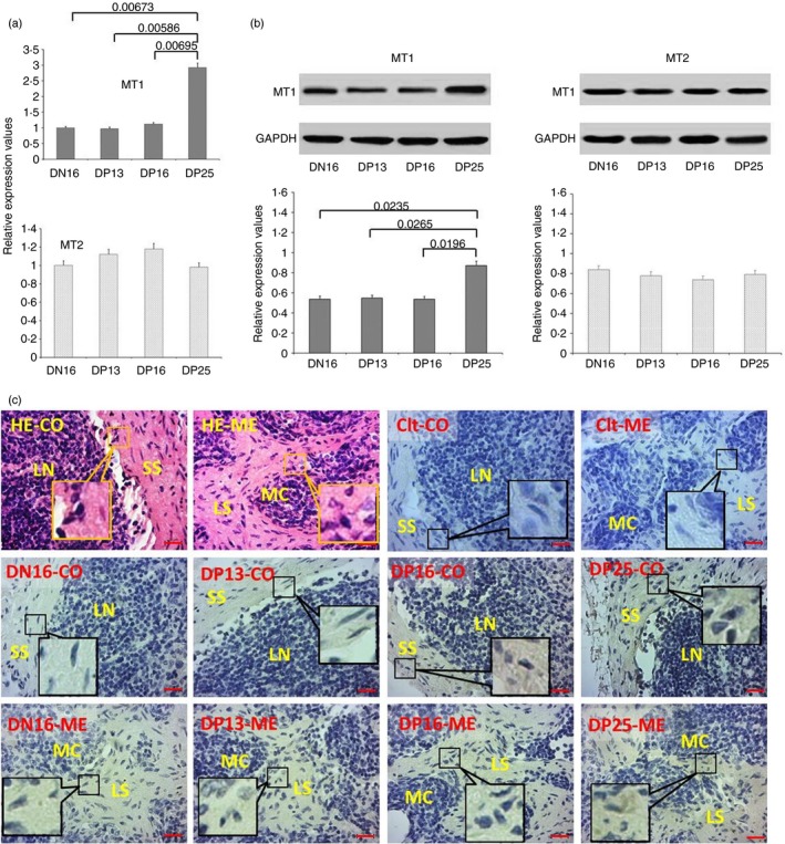
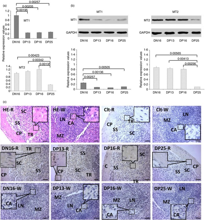
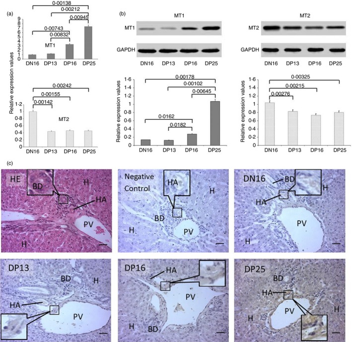
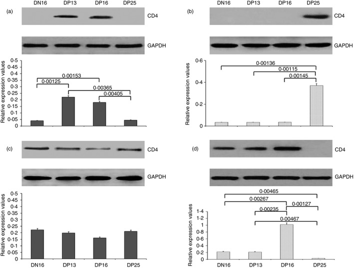
As a pineal gland hormone, melatonin acts through its receptors to modulate the immune system. The immune system is composed of primary and secondary organs, and immune organs are adapted to the presence of the fetal alloantigen during pregnancy. However, it is unclear whether melatonin affects maternal immune organs during early pregnancy in sheep. In this study, the ovine thymus, lymph node, spleen and liver were sampled at day 16 of the oestrous cycle, and at days 13, 16 and 25 of pregnancy. The expression of melatonin receptor 1A (MT1), melatonin receptor 1B (MT2) and cluster of differentiation 4 (CD4) was detected by quantitative real-time polymerase chain reaction, Western blot and immunohistochemistry experiments. Our results showed that during early pregnancy there was an upregulation of MT1 mRNA and protein in the thymus, lymph node and liver, and there was a downregulation in the spleen. The expression of MT2 mRNA and protein was increased in the thymus but decreased in the spleen and liver, and there was no significant change in the lymph node during early pregnancy. CD4 protein was upregulated in the thymus, lymph node and liver, but there were no significant changes in the spleen during early pregnancy. In conclusion, early pregnancy induces tissue-specific expression of MT1, MT2 and CD4, which may be due to the different functions of the thymus, lymph node, spleen and liver. Further, melatonin is involved in immune regulation of the maternal thymus, lymph node, spleen and liver during early pregnancy in sheep.

Keywords: liver; lymph node; melatonin receptor; spleen; thymus.


Figures

Figure 1
Figure 1
Expression of melatonin receptor 1A (MT1) and melatonin receptor 1B (MT2) in thymuses of non‐pregnant and pregnant ewes. (a) Expression values of MT1 and MT2 mRNA in the thymus. (b) Expression of MT1 and MT2 proteins in the thymus. (c) Representative immunohistochemical localization of MT2 protein in the thymuses. The thymus is divided into the cortex (CO) and the medulla (ME). Note: HE = stained by haematoxylin and eosin; Ctl = negative control; DN16 = day 16 of the oestrous cycle; DP13 = day 13 of pregnancy; DP16 = day 16 of pregnancy; DP25 = day 25 of pregnancy; T = thymocyte; ER = epithelial reticular cell; CA = capillary; TC = thymic corpuscle. Scale bar: 20 µm. P < 0·05 indicates significant difference.
Figure 2
Figure 2
Expression of melatonin receptor 1A (MT1) and melatonin receptor 1B (MT2) in lymph nodes of non‐pregnant and pregnant ewes. (a) Expression values of MT1 and MT2 mRNA in the lymph node. (b) Expression of MT1 and MT2 proteins in the lymph node. (c) Representative immunohistochemical localization of MT1 protein in the lymph node. The lymph node is divided into an outer cortex (CO) and an inner medulla (ME). Lymph enters the convex through the subcapsular sinus (SS) and trabeculae (TRs) around the lymphoid nodules (LN) and flows into the medulla through the lymph sinus (LS) around the medullary cord (MC). Note: HE = stained by haematoxylin and eosin; Ctl = negative control; DN16 = day 16 of the oestrous cycle; DP13 = day 13 of pregnancy; DP16 = day 16 of pregnancy; DP25 = day 25 of pregnancy. Scale bar: 20 µm. P < 0·05 indicates significant difference.
Figure 3
Figure 3
Expression of melatonin receptor 1A (MT1) and melatonin receptor 1B (MT2) in the spleens of non‐pregnant and pregnant ewes. (a) Expression values of MT1 and MT2 mRNA in the spleen. (b) Expression of MT1 and MT2 proteins in the spleen. (c) Representative immunohistochemical localization of MT2 protein in the spleen. The spleen is divided into red pulp (R) and white pulp (W), and surrounded by a thickened capsule (CP). Capsules (CPs) with several trabeculae (TRs) project into the substance of the spleen. Note: HE = stained by haematoxylin and eosin; Ctl = negative control; SS = splenic sinuses; SC = splenic cords; MZ = marginal zone; LN = lymphoid nodule; CA = central arteriole; DN16 = day 16 of the oestrous cycle; DP13 = day 13 of pregnancy; DP16 = day 16 of pregnancy; DP25 = day 25 of pregnancy. Scale bar: 50 µm. P < 0·05 indicates significant difference.
Figure 4
Figure 4
Expression of melatonin receptor 1A (MT1) and melatonin receptor 1B (MT2) in the livers of non‐pregnant and pregnant ewes. (a) Expression values of MT1 and MT2 mRNA in the liver. (b) Expression of MT1 and MT2 proteins in the liver. (c) Representative immunohistochemical localization of MT1 protein in the liver. The liver is divided into lobes, and each lobe is made up of hepatic lobules. A portal triad is a component of the hepatic lobule and consists of the proper hepatic artery (HA), hepatic portal vein (PV), small bile ductile (BD) and lymphatic vessels (LV). Note: H = hepatic cell; HE = stained by haematoxylin and eosin; DN16 = day 16 of the oestrous cycle; DP13 = day 13 of pregnancy; DP16 = day 16 of pregnancy; DP25 = day 25 of pregnancy. Scale bar: 50 µm. P < 0·05 indicates significant difference.
Figure 5
Figure 5
Expression of cluster of differentiation 4 (CD4) protein in the thymus, lymph node, spleen and liver. (a) Thymus; (b) lymph node; (c) spleen; (d) liver. DN16 = day 16 of the oestrous cycle; DP13 = day 13 of pregnancy; DP16 = day 16 of pregnancy; DP25 = day 25 of pregnancy. P < 0·05 indicates significant difference.

References

    1. Williams Z. Inducing tolerance to pregnancy. N Engl J Med 2012; 367:1159–61. - PMC - PubMed
    1. Hansen PJ. The immunology of early pregnancy in farm animals. Reprod Domest Anim 2011; 46:18–30. - PubMed
    1. Oliveira LJ, Barreto RS, Perecin F, Mansouri‐Attia N, Pereira FT, Meirelles FV. Modulation of maternal immune system during pregnancy in the cow. Reprod Domest Anim 2012; 47:384–93. - PubMed
    1. Yang L, Zhang LY, Qiao HY, Liu N, Wang YX, Li SJ. Maternal immune regulation by conceptus during early pregnancy in the bovine. Asian J Anim Vet Adv 2014; 9:610–20.
    1. Parker GA, Papenfuss TL. Chapter 10 ‐ Immune system In: Atlas of Histology of the Juvenile Rat. New York: Academic Press, 2016: 293–347.

Publication types

MeSH terms

Associated data