. 2020 Mar;245(6):522-531.
doi: 10.1177/1535370220906541. Epub 2020 Feb 13.

Salidroside promotes sciatic nerve regeneration following combined application epimysium conduit and Schwann cells in rats

Affiliations

Salidroside promotes sciatic nerve regeneration following combined application epimysium conduit and Schwann cells in rats

Jiaqi Li et al. Exp Biol Med (Maywood). 2020 Mar.

Abstract

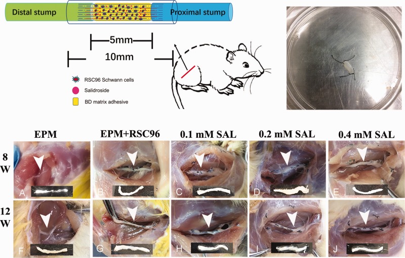
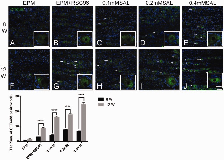
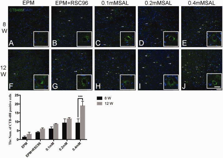
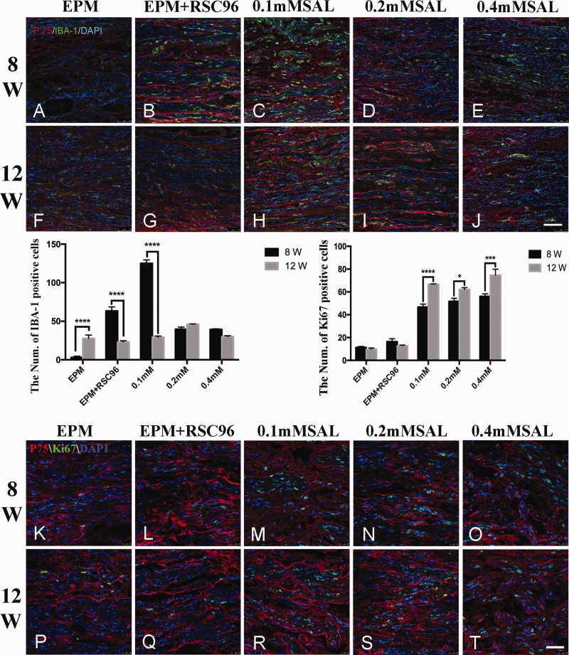
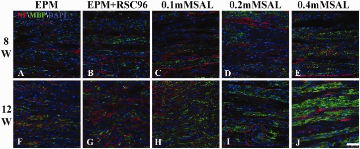
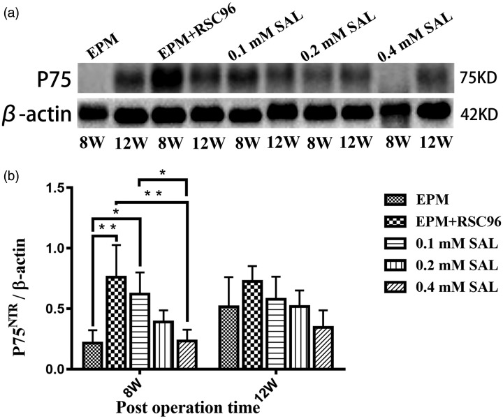
Schwann cell and nerve conduit are crucial for nerve regeneration and re-myelination after peripheral nerves injury. To investigate the effects of Salidroside on autogenous epimysium conduit mixed with BD matrigel and RSC96 Schwann cells on an animal model with 5 mm sciatic nerve defect injury in rat, motor function, muscle reinnervation, immunohistochemical staining, retrograded tracing and Western blot were used in this study. The results showed that Salidroside enhanced the compound effects of epimysium conduit mixed with BD matrigel and RSC96 Schwann cells to improve the sciatic functional index and the gastrocnemius muscle weight ratio, which were better than EPM group at 8 weeks and 12 weeks post operation. Immunofluorescence and Western blot results of P75NTR showed that Salidroside improved the sciatic nerve regeneration, and retrograded tracing of CTB-Alexa 488 also supported that Salidroside was better to promote CTB tracer transporting from the distal nerve defect to the ipsilateral dorsal root ganglion and ventral horn of L3-L5 spinal cord on post-operation 8 weeks and 12 weeks. Our results demonstrated that Salidroside improved the effect of autogenous epimysium conduit mixed with BD matrigel and RSC96 Schwann cells on sciatic nerve regeneration in our study.

Impact statement: Peripheral nerve injury and regeneration remain a major challenge. Although nerve conduit and Schwann cells have been used to study the nerve regeneration, our results demonstrated that Salidroside improved the regenerative effect in a rat model with sciatic nerve injury model, following a combined application of autogenous epimysium conduit mixed with Schwann cells. Different concentrations of Salidroside combining autogenous epimysium conduit and Schwann cells were applied to compare the epimysium conduit group and the epimysium conduit combining Schwann cells group. Based on the results of motor function and muscle reinnervation evaluation, as well as neuronal tracing and expression of P75NTR, our study for the first time suggests that Salidroside may improve the regeneration effect on the sciatic nerve following a combined application of epimysium conduit and RSC96 Schwann cells in rats.

Keywords: RSC96 Schwann cells; Salidroside; autologous epimysium conduit; combination application; nerve regeneration; sciatic nerve injury.


Figures

Figure 1.
Figure 1.
Diagram of animal surgery for sciatic nerve injury model and gross observation of regenerative sciatic nerve. An autologous epimysium conduit was performed to bridge both stumps of the nerve defect. BD matrigel, Schwann cells, and Salidroside (SAL) mixture were injected inside the epimysium conduit during operation (top left). The epimysium slice were obtain from the right external oblique and fixed with 6–0 nylon sutures for subsequent epimysium conduit preparation (top right). In post-operation 8 weeks, poor bridge of the stumps was observed in the sham group (a), while the sciatic nerve injury in other groups was completely connected together by regenerative tissue (b–e); in post-operation 12 weeks (f–j), the regenerative nerves of all the groups were relatively coarser than before, and the 0.2 mM Salidroside mixture group (i) and 0.4 mM Salidroside mixture group (j) were better regenerative. Arrowhead showed the epimysium conduit bridge the sciatic nerve injury, and below was the representative segment of regeneration sciatic nerve tissue. (A color version of this figure is available in the online journal.)
Figure 2.
Figure 2.
Retrograde labeling of CTB-Alexa 488 (green) in the dorsal root ganglion. In post-operation 8 weeks, no obvious CTB-Alexa 488-positive granules in the sham group were observed (a), while other groups (b–e) were only relatively obvious than the sham group; In post-operation 12 weeks, the CTB-Alexa 488-positive granules in the different groups increased than post-operation 8 weeks, and the CTB-Alexa 488-positive granules in the Salidroside (SAL) mixture groups (h–j) were relatively more than the sham group and BDSC group (f–g), especially the 0.4 mM Salidroside mixture group (j). White arrow showed the CTB-Alexa 488-labeled neuron. Histogram showed the count of CTB-Alexa 488-labeled neurons in the dorsal root ganglion. n = 4; Bar = 75 μm. (Enlarged view bar = 30 μm) ****P < 0.0001. (A color version of this figure is available in the online journal.)
Figure 3.
Figure 3.
Retrograde labeling of CTB-Alexa 488 (green) in the anterior horn of L3–L5 spinal cord. In post-operation 8 weeks, CTB-labeled neurons were absent in the right ventral horn in sham group and BDSC group (a–b), but only a few CTB-labeled neurons were observed in the Salidroside (SAL) mixture groups (c–e). In post-operation 12 weeks, CTB-labeled neurons presented in all the groups (f–j), and the 0.4 mM Salidroside mixture group (j) was relatively more than other groups. White arrow showed the CTB-Alexa 488-labeled neuron. Histogram showed the count of CTB-Alexa 488-labeled neurons in the anterior horn of L3–L5. n = 4; Bar = 75 μm. (Enlarged view bar = 30 μm) ***P < 0.001. (A color version of this figure is available in the online journal.)
Figure 4.
Figure 4.
Immunofluorescence staining of P75NTR, IBA-1, and Ki67 in regenerated sciatic nerve. In post-operation 8 weeks, the regenerative nerve fibers were disordered arrangement and only a few P75NTR was observed in the sham group (a). Compared with the sham group, the regenerative nerve fibers in the other groups arranged relatively regular (b–e), P75NTR and Iba-1 immunoreactivities were also observed. In post-operation 12 weeks, the regenerative nerve fibers in all the groups (f–j) obviously improved than before, P75NTR immunoreactivities in different Salidroside (SAL) mixture groups were better than sham group (f) and BDSC group (g), and the expression of Iba-1 in 0.2 mM Salidroside mixture group (i) was also lower than other group. Histograms showed the count of IBA-1-positive cells and Ki67-positive cells in regenerative sciatic nerve. Ki67-positive cells were observed in all groups at post-operation 8 weeks and 12 weeks, but there were more obvious in the Salidroside mixture groups with different concentrations than EPM group and EPM + RSC96 group (k–t). n = 5; Bar = 100 μm. (A color version of this figure is available in the online journal.)
Figure 5.
Figure 5.
Immunofluorescence staining of Neurofilament and MBP in regenerated sciatic nerve. At 8 weeks post-operation, the neurofilament (red) and MBP- (green) positive regenerative tissues longitudinally distributed along the sciatic nerve, and their expression in 0.4 mM Salidroside mixture group was more obvious than other groups, while the EPM group showed fainter expression. At 12 weeks post-operation, the regenerative nerve tissues in all groups seemed to be arranged relatively regular and more intensive than those at 8 weeks post-operation, especially the neurofilament in 0.4 mM Salidroside mixture group was observed more expression than others and better alignment. n = 5; Bar = 50 μm. (A color version of this figure is available in the online journal.)
Figure 6.
Figure 6.
Western blot of P75NTR in the regenerative sciatic nerve. The expression of P75NTR in the sham group was not obvious in post-operation 8 weeks; all the other Salidroside (SAL) mixture groups were higher than sham group. In post-operation 12 weeks, there were no significant difference in the expression of P75NTR in all the groups (n = 5). *P < 0.05, **P < 0.01.

Similar articles

Cited by

References

    1. Ciaramitaro P, Mondelli M, Logullo F, Grimaldi S, Battiston B, Sard A, Scarinzi C, Migliaretti G, Faccani G, Cocito D; Italian network for traumatic N. Traumatic peripheral nerve injuries: epidemiological findings, neuropathic pain and quality of life in 158 patients. J Peripher Nerv Syst 2010; 15:120–7 - PubMed
    1. Chrzaszcz P, Derbisz K, Suszynski K, Miodonski J, Trybulski R, Lewin-Kowalik J, Marcol W. Application of peripheral nerve conduits in clinical practice: a literature review. Neurol Neurochir Pol 2018; 52:427–35 - PubMed
    1. Han GH, Peng J, Liu P, Ding X, Wei S, Lu S, Wang Y. Therapeutic strategies for peripheral nerve injury: decellularized nerve conduits and Schwann cell transplantation. Neural Regen Res 2019; 14:1343–51 - PMC - PubMed
    1. de Luca AC, Lacour SP, Raffoul W, di Summa PG. Extracellular matrix components in peripheral nerve repair: how to affect neural cellular response and nerve regeneration? Neural Regen Res 2014; 9:1943–8 - PMC - PubMed
    1. Jiang Z, Song Y, Qiao J, Yang Y, Zhang W, Liu W, Han B. Rat sciatic nerve regeneration across a 10-mm defect bridged by a chitin/CM-chitosan artificial nerve graft. Int J Biol Macromol 2019; 129:997–1005 - PubMed

Publication types