

Computational Biomechanical Analysis of Postoperative Calcaneal Fractures with Different Placement of the Sustentaculum Screw
- PMID: 32053281
- PMCID: PMC7189067
- DOI: 10.1111/os.12541
Computational Biomechanical Analysis of Postoperative Calcaneal Fractures with Different Placement of the Sustentaculum Screw
Abstract
Objective: To evaluate the computational biomechanical analysis of intra-articular calcaneal fractures with different fixation status of the sustentaculum plate screw, when the finite element modeling of calcaneal fractures were fixed by the lateral locking plate.
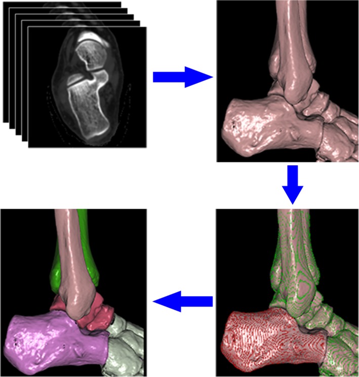
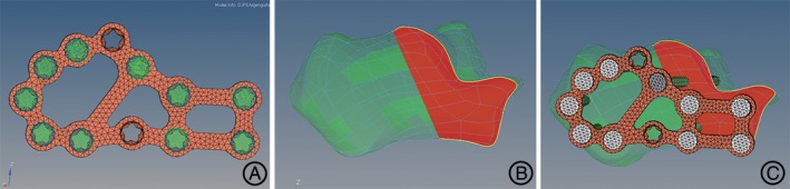
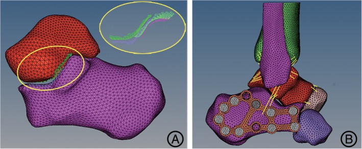
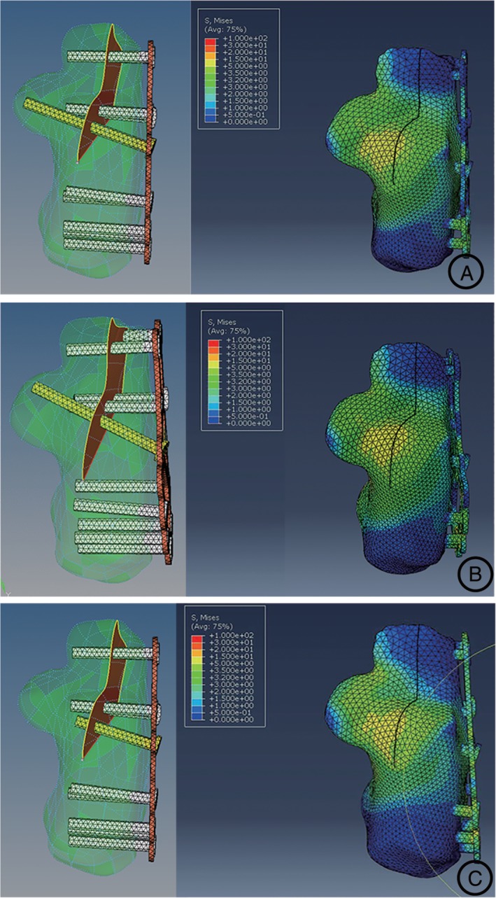
Methods: The normal right foot of a male (age: 36 years; height: 174 cm; body weight: 65 kg) was scanned by the CT scanner. As the computational biomechanical study, the three-dimensional finite element model of the simplified Sanders type-II calcaneal fracture was built. Fixation with the lateral calcaneal locking plate and screws was simulated using a finite element software package according to clinical operation. According to the different placement of the sustentaculum plate screw, the models were categorized as the accurate fixation group, marginal fixation group, and non-fixation group. The loading of 650 N with the vertical axial compression was applied to simulate the standing phase with single foot. The Von Mises stress distribution, maximal displacement, and contact area of the subtalar joint were analyzed among three groups.
Results: The pressure distribution of the subtalar joint facet was inhomogeneous. The stress concentration of the calcaneus was located at the medial zone of the posterior subtalar joint facet. The peak Von Mises stress distribution in three groups was similar at the subtalar joint facet of 4.9 MPa, 5.1 MPa, and 5.4 MPa. In the accurate fixation group, the contact area on the posterior articular facet was 277.1 mm2 ; the maximal displacement was 0.18 mm. The contact area of the marginal fixation group was 265.3 mm2 on the posterior facet, where the maximal displacement was 0.23 mm. In the non-fixation group, the contact area was 253.8 mm2 ; the maximal displacement was 0.25 mm. There was a slight change in the contact area of the subtalar joint and no prominent displacement of the calcaneus could be detected among the three groups.
Conclusions: The biomechanical results, including the peak stress distribution, contact area, and maximal displacement of subtalar joint, were similar whether the screw is placed exactly within the sustentaculum tali or not, when the calcaneal fractures were fixed by the lateral locking plate. The sustentaculum plate screw had less effect on the biomechanical performance of the calcaneus.
Keywords: Calcaneus; Finite element analysis; Fracture fixation, Internal; Intra-articular fractures.
© 2019 The Authors. Orthopaedic Surgery published by Chinese Orthopaedic Association and John Wiley & Sons Australia, Ltd.
Figures
References
-
- Potter MQ, Nunley JA. Long‐term functional outcomes after operative treatment for intra‐articular fractures of the calcaneus. J Bone Joint Surg Am, 2009, 91: 1854–1860. - PubMed
-
- Guerado E, Bertrand ML, Cano JR. Management of calcaneal fractures: what have we learnt over the years? Injury, 2012, 43: 1640–1650. - PubMed
-
- Swanson SA, Clare MP, Sanders RW. Management of intra‐articular fractures of the calcaneus. Foot Ankle Clin, 2008, 13: 659–678. - PubMed
-
- Buckley R, Tough S, Mccormack R, et al Operative compared with nonoperative treatment of displaced intra‐articular calcaneal fractures: a prospective, randomized, controlled multicenter trial. J Bone Joint Surg Am, 2002, 84: 1733–1744. - PubMed
-
- Basile A. Operative versus nonoperative treatment of displaced intra‐articular calcaneal fractures in elderly patients. J Foot Ankle Surg, 2010, 49: 25–32. - PubMed
MeSH terms
Grants and funding
LinkOut - more resources
Full Text Sources
Medical
Research Materials
