

Time Course of Lung Changes at Chest CT during Recovery from Coronavirus Disease 2019 (COVID-19)
- PMID: 32053470
- PMCID: PMC7233367
- DOI: 10.1148/radiol.2020200370
Time Course of Lung Changes at Chest CT during Recovery from Coronavirus Disease 2019 (COVID-19)
Abstract
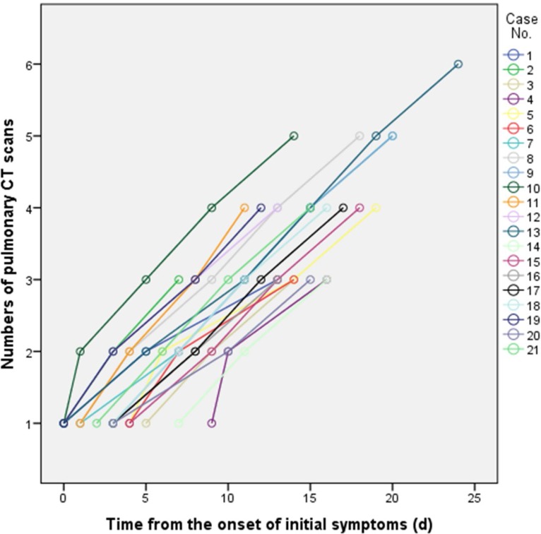
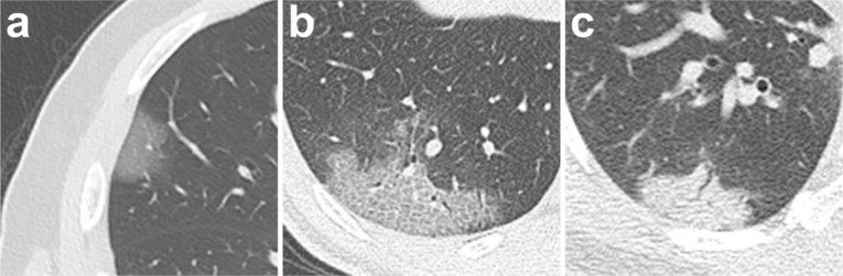
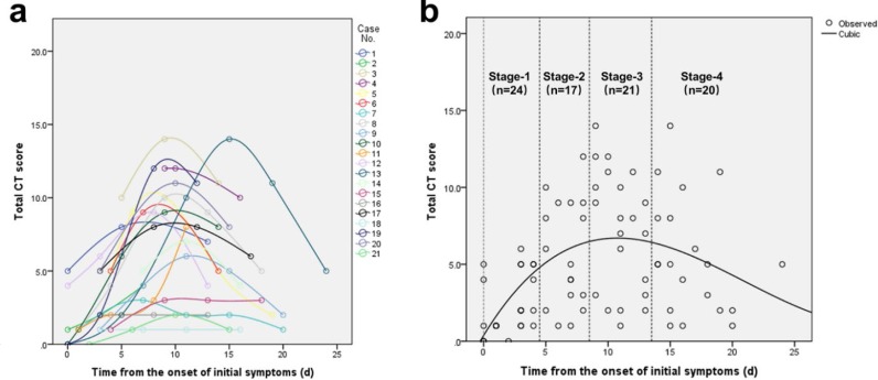
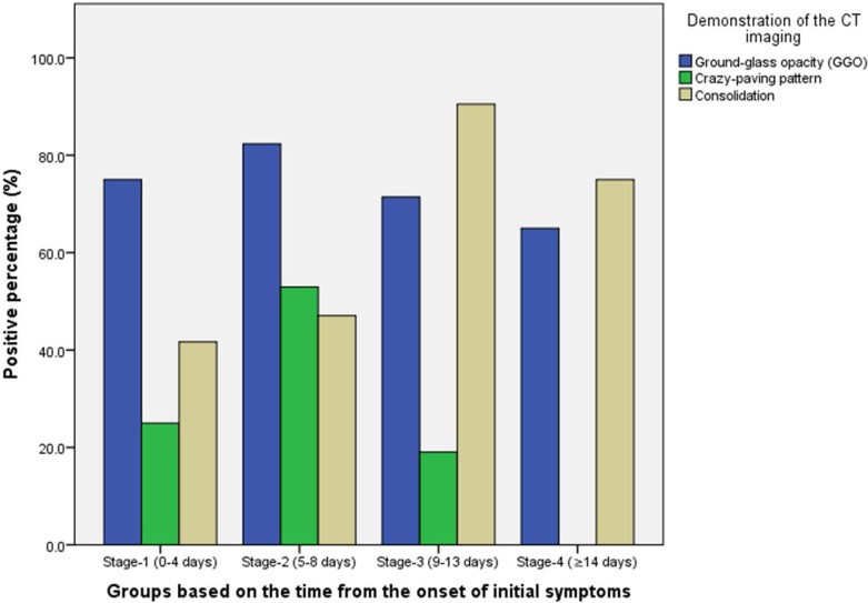
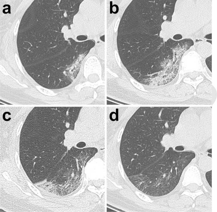
Background Chest CT is used to assess the severity of lung involvement in coronavirus disease 2019 (COVID-19). Purpose To determine the changes in chest CT findings associated with COVID-19 from initial diagnosis until patient recovery. Materials and Methods This retrospective review included patients with real-time polymerase chain reaction-confirmed COVID-19 who presented between January 12, 2020, and February 6, 2020. Patients with severe respiratory distress and/or oxygen requirement at any time during the disease course were excluded. Repeat chest CT was performed at approximately 4-day intervals. Each of the five lung lobes was visually scored on a scale of 0 to 5, with 0 indicating no involvement and 5 indicating more than 75% involvement. The total CT score was determined as the sum of lung involvement, ranging from 0 (no involvement) to 25 (maximum involvement). Results Twenty-one patients (six men and 15 women aged 25-63 years) with confirmed COVID-19 were evaluated. A total of 82 chest CT scans were obtained in these patients, with a mean interval (±standard deviation) of 4 days ± 1 (range, 1-8 days). All patients were discharged after a mean hospitalization period of 17 days ± 4 (range, 11-26 days). Maximum lung involved peaked at approximately 10 days (with a calculated total CT score of 6) from the onset of initial symptoms (R2 = 0.25, P < .001). Based on quartiles of chest CT scans from day 0 to day 26 involvement, four stages of lung CT findings were defined. CT scans obtained in stage 1 (0-4 days) showed ground-glass opacities (18 of 24 scans [75%]), with a mean total CT score of 2 ± 2; scans obtained in stage 2 (5-8 days) showed an increase in both the crazy-paving pattern (nine of 17 scans [53%]) and total CT score (mean, 6 ± 4; P = .002); scans obtained in stage 3 (9-13 days) showed consolidation (19 of 21 scans [91%]) and a peak in the total CT score (mean, 7 ± 4); and scans obtained in stage 4 (≥14 days) showed gradual resolution of consolidation (15 of 20 scans [75%]) and a decrease in the total CT score (mean, 6 ± 4) without crazy-paving pattern. Conclusion In patients recovering from coronavirus disease 2019 (without severe respiratory distress during the disease course), lung abnormalities on chest CT scans showed greatest severity approximately 10 days after initial onset of symptoms. © RSNA, 2020.
Figures
Comment in
-
Essentials for Radiologists on COVID-19: An Update-Radiology Scientific Expert Panel.Radiology. 2020 Aug;296(2):E113-E114. doi: 10.1148/radiol.2020200527. Epub 2020 Feb 27. Radiology. 2020. PMID: 32105562 Free PMC article. No abstract available.
-
Patients with RT-PCR-confirmed COVID-19 and Normal Chest CT.Radiology. 2020 May;295(2):E3. doi: 10.1148/radiol.2020200702. Epub 2020 Mar 6. Radiology. 2020. PMID: 32142398 Free PMC article. No abstract available.
-
Can Lung US Help Critical Care Clinicians in the Early Diagnosis of Novel Coronavirus (COVID-19) Pneumonia?Radiology. 2020 Jun;295(3):E6. doi: 10.1148/radiol.2020200847. Epub 2020 Mar 13. Radiology. 2020. PMID: 32167853 Free PMC article. No abstract available.
-
Chest Radiograph Findings in Asymptomatic and Minimally Symptomatic Quarantined Patients in Codogno, Italy during COVID-19 Pandemic.Radiology. 2020 Jun;295(3):E7. doi: 10.1148/radiol.2020201102. Epub 2020 Mar 27. Radiology. 2020. PMID: 32216718 Free PMC article. No abstract available.
-
Letter to the Editor: Update on the followed-up CT exam of the first CoVID-19 pneumonia in Taiwan.J Formos Med Assoc. 2020 Jun;119(6):1121-1122. doi: 10.1016/j.jfma.2020.04.012. Epub 2020 Apr 20. J Formos Med Assoc. 2020. PMID: 32334915 Free PMC article. No abstract available.
-
Covid-19 Interstitial Pneumonia: Histological and Immunohistochemical Features on Cryobiopsies.Respiration. 2021;100(6):488-498. doi: 10.1159/000514822. Epub 2021 Mar 16. Respiration. 2021. PMID: 33725700 Free PMC article.
References
-
- Huang C, Wang Y, Li X, Ren L, Zhao J, Hu Y, Zhang L, Fan G, Xu J, Gu X, Cheng Z, Yu T, Xia J, Wei Y, Wu W, Xie X, Yin W, Li H, Liu M, Xiao Y, Gao H, Guo L, Xie J, Wang G, Jiang R, Gao Z, Jin Q, Wang J, Cao B. Clinical features of patients infected with 2019 novel coronavirus in Wuhan, China. Lancet 2020. doi: 10.1016/S0140-6736(20)30183-5 - PMC - PubMed
-
- Li Q, Guan X, Wu P, Wang X, Zhou L, Tong Y, Ren R, Leung KSM, Lau EHY, Wong JY, Xing X, Xiang N, Wu Y, Li C, Chen Q, Li D, Liu T, Zhao J, Li M, Tu W, Chen C, Jin L, Yang R, Wang Q, Zhou S, Wang R, Liu H, Luo Y, Liu Y, Shao G, Li H, Tao Z, Yang Y, Deng Z, Liu B, Ma Z, Zhang Y, Shi G, Lam TTY, Wu JTK, Gao GF, Cowling BJ, Yang B, Leung GM, Feng Z. Early Transmission Dynamics in Wuhan, China, of Novel Coronavirus-Infected Pneumonia. The New England journal of medicine 2020. doi: 10.1056/NEJMoa2001316 - PMC - PubMed
-
- Zhu N, Zhang D, Wang W, Li X, Yang B, Song J, Zhao X, Huang B, Shi W, Lu R, Niu P, Zhan F, Ma X, Wang D, Xu W, Wu G, Gao GF, Tan W, China Novel Coronavirus I, Research T. A Novel Coronavirus from Patients with Pneumonia in China, 2019. The New England journal of medicine 2020. doi: 10.1056/NEJMoa2001017 - PMC - PubMed
-
- World Health Organization . Novel coronavirus (2019-nCoV). Situation report 22. Geneva, Switzerland: World Health Organization; 2020. https://www.who.int/docs/default-source/coronaviruse/situation-reports/2... Published on February 11, 2020.
MeSH terms
LinkOut - more resources
Full Text Sources
Other Literature Sources
Medical
