

Optical Coherence Tomography Angiography in Glaucoma
- PMID: 32053551
- PMCID: PMC7117982
- DOI: 10.1097/IJG.0000000000001463
Optical Coherence Tomography Angiography in Glaucoma
Abstract
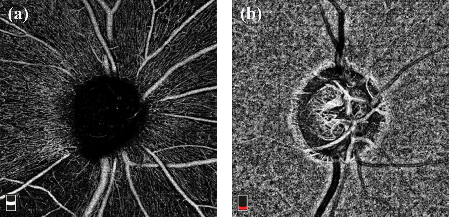
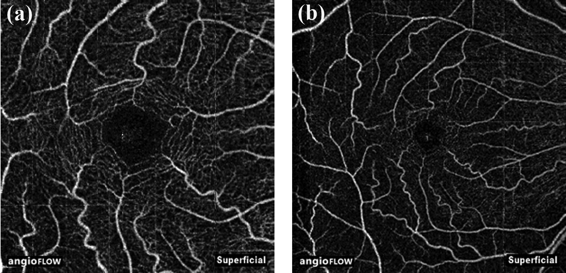
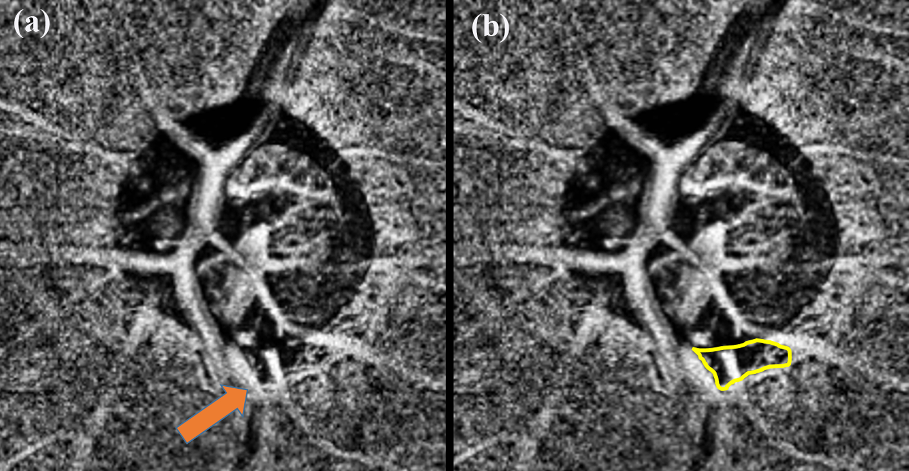
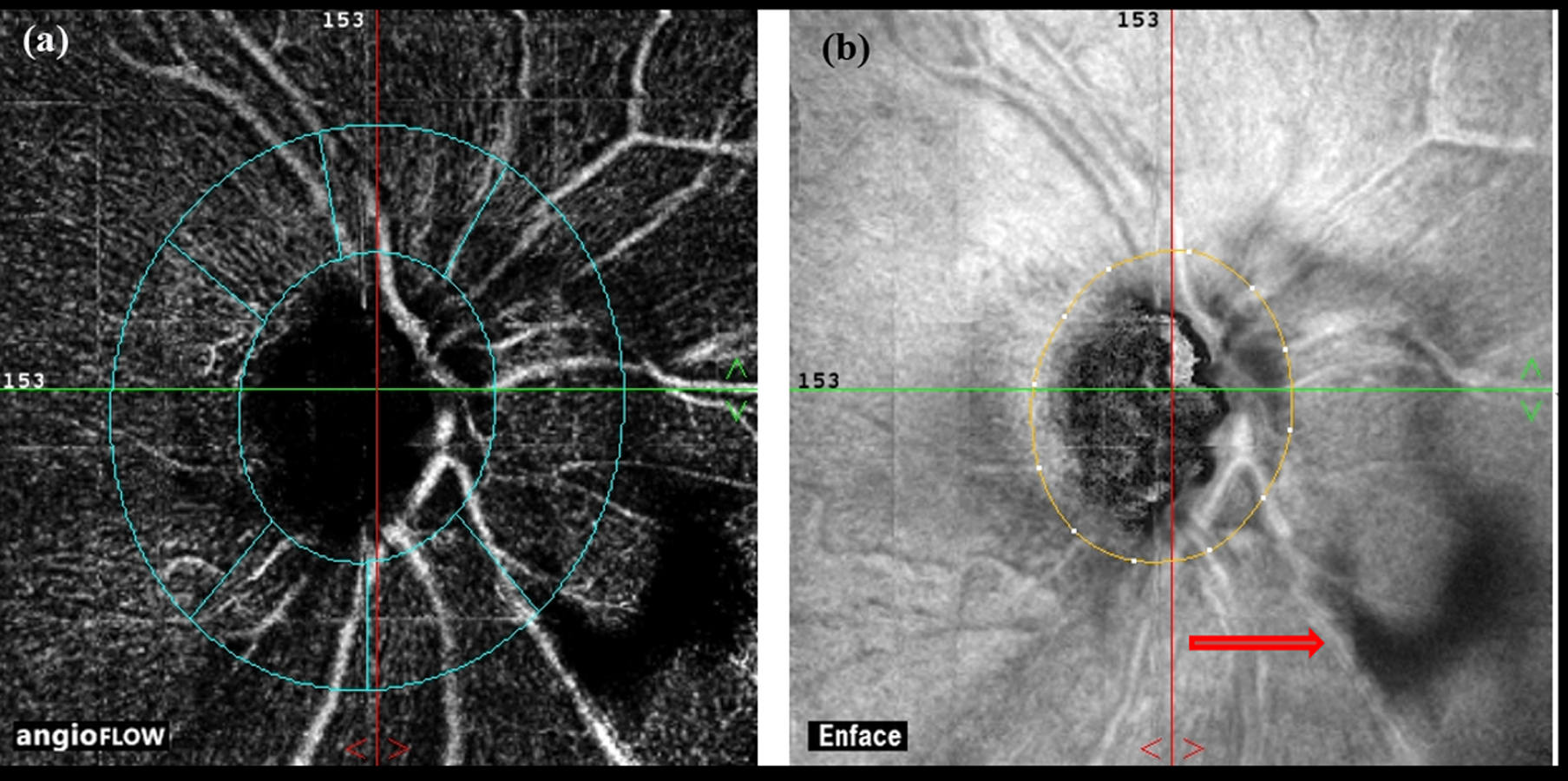
Optical coherence tomography angiography (OCTA) is a relatively new, noninvasive, dye-free imaging modality that provides a qualitative and quantitative assessment of the vasculature in the retina and optic nerve head. OCTA also enables visualization of the choriocapillaris, but only in areas of parapapillary atrophy. With OCTA, the movement of red blood cells is used as a contrast to delineate blood vessels from static tissues. The features seen with OCTA in eyes with glaucoma are reduction in the superficial vessel density in the peripapillary and macular areas, and complete loss of choriocapillaris in localized regions of parapapillary atrophy (called deep-layer microvascular dropout). These OCTA changes correlate well topographically with the functional changes seen on visual field examination and structural changes seen on optical coherence tomography (OCT) (ie, parapapillary retinal nerve fiber layer changes and inner retinal layer thickness changes at macula). The OCTA measurements also have acceptable test-retest variability and well differentiate glaucomatous from normal eyes. OCTA measurements can be affected by various subject-related, eye-related, and disease-related factors. Vessel density reduction on OCTA reaches a base level (floor) at a more advanced disease stage than the structural changes on OCT and therefore has the potential to monitor progression in eyes with advanced glaucomatous damage. OCTA also adds information about glaucoma patients at risk of faster progression. OCTA, therefore, complements visual field and OCT examinations to diagnose glaucoma, detect progression, and assess risk of progression.
Figures


References
-
- Fechtner RD, Weinreb RN. Mechanisms of optic nerve damage in primary open angle glaucoma. Surv Ophthalmol 1994;39:23–42. - PubMed
-
- Sommer A, Tielsch JM, Katz J, et al. Relationship between intraocular pressure and primary open angle glaucoma among white and black Americans. The Baltimore Eye Survey. Arch Ophthalmol 1991;109:1090–5. - PubMed
