

The Microbiome of the Meibum and Ocular Surface in Healthy Subjects
- PMID: 32053729
- PMCID: PMC7326502
- DOI: 10.1167/iovs.61.2.18
The Microbiome of the Meibum and Ocular Surface in Healthy Subjects
Abstract
Purpose: The purpose of this study was to investigate the microbiome in the meibum, conjunctival sac, and eyelid skin in young and elderly healthy subjects, and analyze the effect that age, sex, and region have on microbiome composition.
Methods: This study involved 36 healthy subjects (young-age subjects: 9 men/9 women, age range: 20-35 years; elderly age subjects: 9 men/9 women, age range: 60-70 years). In all subjects, lower-eyelid meibum, lower conjunctival sac, and lower-eyelid skin specimens were collected from one eye, and then stored at -20°C. Taxonomic composition of the microbiome was obtained via 16S rRNA gene sequencing, and then analyzed.
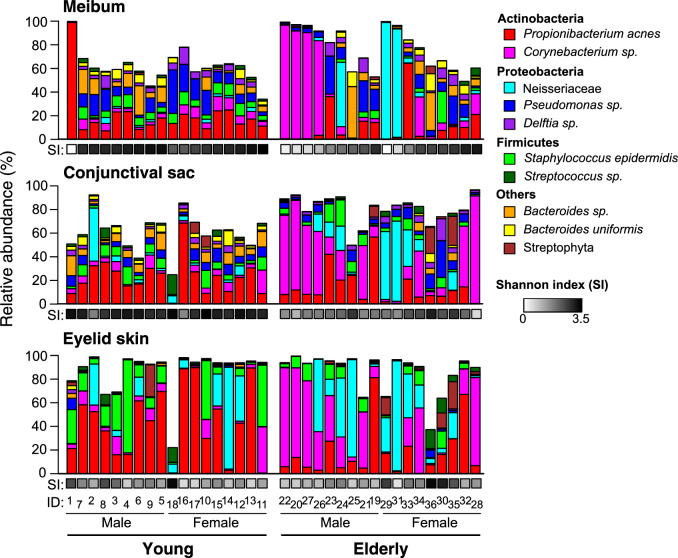
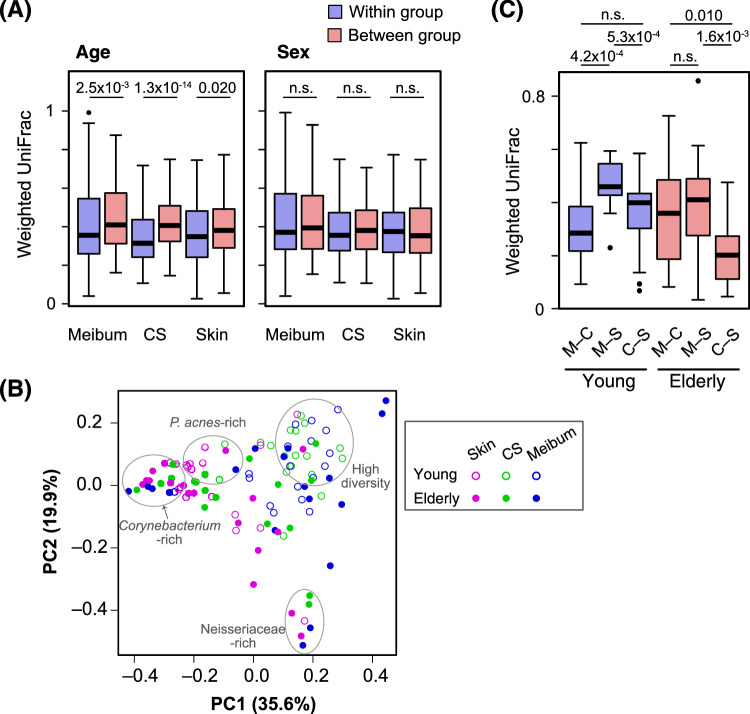
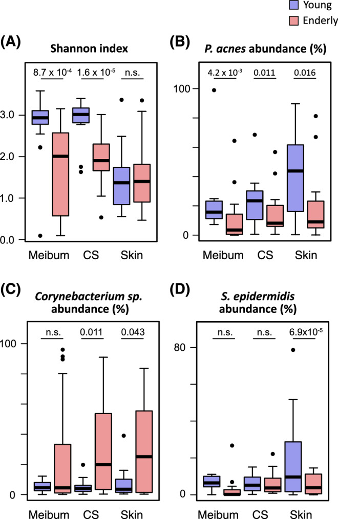
Results: The meibum microbiome showed a high α-diversity (within-community diversity), particularly in the young subjects. However, in approximately 30% of the elderly subjects, a low-diversity microbiome dominated by Corynebacterium sp. or Neisseriaceae was observed. In the young subjects, the microbiome of the meibum resembled that of the conjunctival-sac, yet in the elderly subjects, the microbiome of the conjunctival-sac became more similar to that of the eyelid skin. The eyelid-skin microbiome was relatively simple, and was typically dominated by Propionibacterium acnes in the young subjects, or by Corynebacterium sp. or Neisseriaceae in the elderly subjects. In both age groups, no significant difference was seen between the men and women in regard to the meibum, conjunctival-sac, and eyelid-skin microbiome.
Conclusions: Our findings confirmed that the meibum of healthy adult-age subjects harbors highly diverse microbiota, and revealed that the meibum microbiome, especially the decrease of its diversity, alters with aging and may affect the homeostasis of the ocular surface.
Conflict of interest statement
Disclosure:
Figures
References
-
- Suzuki T, Teramukai S, Kinoshita S. Meibomian glands and ocular surface inflammation. Ocul Surf. 2015; 13: 133–149. - PubMed
-
- Nicolaides N, Kaitaranta JK, Rawdah TN, Macy JI, Boswell FM 3rd, Smith RE. Meibomian gland studies: comparison of steer and human lipids. Invest Ophthalmol Vis Sci. 1981; 20: 522–536. - PubMed
-
- Bron AJ, Benjamin L, Snibson GR. Meibomian gland disease. Classification and grading of lid changes. Eye (Lond). 1991; 5(Pt 4): 395–411. - PubMed
-
- Montes-Mico R. Role of the tear film in the optical quality of the human eye. J Cataract Refract Surg. 2007; 33: 1631–1635. - PubMed
Publication types
MeSH terms
LinkOut - more resources
Full Text Sources
