. 2020 Mar 24;118(6):1292-1300.
doi: 10.1016/j.bpj.2019.12.040. Epub 2020 Jan 28.

Cell-Derived Plasma Membrane Vesicles Are Permeable to Hydrophilic Macromolecules

Affiliations

Cell-Derived Plasma Membrane Vesicles Are Permeable to Hydrophilic Macromolecules

Allison D Skinkle et al. Biophys J. .

Abstract

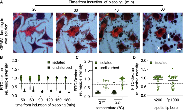
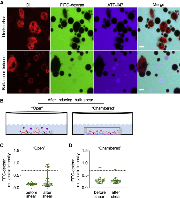
Giant plasma membrane vesicles (GPMVs) are a widely used experimental platform for biochemical and biophysical analysis of isolated mammalian plasma membranes (PMs). A core advantage of these vesicles is that they maintain the native lipid and protein diversity of the PM while affording the experimental flexibility of synthetic giant vesicles. In addition to fundamental investigations of PM structure and composition, GPMVs have been used to evaluate the binding of proteins and small molecules to cell-derived membranes and the permeation of drug-like molecules through them. An important assumption of such experiments is that GPMVs are sealed, i.e., that permeation occurs by diffusion through the hydrophobic core rather than through hydrophilic pores. Here, we demonstrate that this assumption is often incorrect. We find that most GPMVs isolated using standard preparations are passively permeable to various hydrophilic solutes as large as 40 kDa, in contrast to synthetic giant unilamellar vesicles. We attribute this leakiness to stable, relatively large, and heterogeneous pores formed by rupture of vesicles from cells. Finally, we identify preparation conditions that minimize poration and allow evaluation of sealed GPMVs. These unexpected observations of GPMV poration are important for interpreting experiments utilizing GPMVs as PM models, particularly for drug permeation and membrane asymmetry.


Figures

Figure 1
Figure 1
Large solutes permeate GPMVs. (A) Isolated GPMVs exposed to FITC-modified 3-kDa dextran (FITC-dextran) and Alexa-Fluor-modified ATP analog (ATP-647) exhibit accumulation of probes in the intravesicular space of some vesicles. (B) In contrast, GUVs are not permeable to the same probes. This effect is quantified in (C), in which points represent relative intensities of individual vesicles in a given preparation. A robust population of permeable GPMVs (defined by a relative intensity >0.7; see Materials and Methods) is observed, whereas almost all GUVs remain sealed. (D) Permeable GPMVs fill with both probes proportionally, and in all standard preparations two populations emerge: sealed and permeable vesicles. (E) Although the percentage of permeable vesicles varies considerably in repeat preparations, on average, around 65% of isolated GPMVs are permeable. Here, each data point represents the percentage of permeable vesicles in a given preparation of GPMVs. To see this figure in color, go online.
Figure 2
Figure 2
Large solutes permeate GPMVs rapidly. (A) Time-lapse imaging of GPMVs exposed to hydrophilic probes revealed that vesicles fill with solutes in approximately 1 min. (B) Although FITC-dextran signal quickly levels out after this time, ATP-647 signal continues to accumulate, likely through interactions with ATP-binding proteins within vesicles. Dotted lines indicate time to vesicle permeation (reaching a relative vesicle intensity of 0.7), as calculated via one-phase exponential association kinetics. Time to filling is 55 s for FITC-dextran and 49 s for ATP-647. Scale bars, 5 μm. rel., relative. To see this figure in color, go online.
Figure 3
Figure 3
Large solutes permeate GPMVs via stable pores. When exposed to fluorescent dextrans with different molecular weights, GPMV permeation showed a clear size dependence. (A) Some vesicles that were permeable to 3-kDa dextran completely excluded 60-kDa dextran. (B) Dextrans with lower molecular weights permeated GPMVs with decreasing frequency up to 60 kDa, suggesting the existence of pores of heterogeneous sizes. Each data point represents a population of GPMVs from the same preparation incubated with differently sized dextrans. (C) Vesicles that were prepared in the presence of FITC-dextran only were then exposed to ATP-647 30 min after their isolation. Vesicles that were permeable to FITC-dextran at isolation were also permeable to ATP-647 after 30 min, suggesting that pores remain stable for extended periods. Scale bars, 10 μm. To see this figure in color, go online.
Figure 4
Figure 4
GPMVs become permeable upon isolation independent of temperature cycling or pipetting stress. (A) Time-lapse of cells during GPMV production indicates that GPMVs are not permeable immediately upon their budding or during their growth. Both cell-attached (x) and detached (arrow) vesicles remained impermeable to both fluorescent probes. (B) GPMVs isolated from cells were compared with those that were left undisturbed alongside the cells they formed from over the course of 2.5 h. Whereas undisturbed GPMVs remained impermeable thoughout the time course, the isolated samples had a distribution of permeable and impermeable vesicles that remained consistent over time. (C) Keeping temperature constant between formation and isolation by isolating GPMVs at 37°C as opposed to 22°C did not affect the permeation induced by isolation, suggesting that temperature changes during GPMV handling were not responsible for permeation. (D) Changing shear strain by pipetting with different pipette tip bores did not affect the population of permeable vesicles, suggesting that pipetting is not responsible for eliciting permeation. Asterisks indicate that the pipette tip was cut to increase bore. All results from (B)–(D) were repeated in the presence of ATP-647 as demonstrated in the Supporting Material. Scale bar, 10 μm. rel., relative. To see this figure in color, go online.
Figure 5
Figure 5
GPMVs become permeable during shear-induced detachment from cells. (A) When a plate of cells forming GPMVs is jostled, shear stress from movement of the bulk solution is induced, and a previously sealed population of GPMVs becomes permeable. (B) Schematic. GPMVs forming in this manner (open configuration) become permeable because of shear flow, likely by rupture from cells. In contrast, GPMV formation can be induced in a sealed chamber (“chambered” configuration), which eliminates shear forces from movement of the bulk solution. Chambered GPMVs remain sealed despite jostling of the plate. (C) GPMVs forming in the open configuration became permeable upon generating shear from movement of the bulk solution. (D) In contrast, GPMVs forming in the chambered configuration remain impermeable regardless of bulk fluid movement. In (C) and (D), data points represent individual vesicles in a preparation of GPMVs. Scale bar, 10 μm. rel., relative. To see this figure in color, go online.
Figure 6
Figure 6
GPMVs formed in shear-protected conditions remain sealed. (A) GPMVs formed in a shear-protected chamber can be separated from cells by inverting the chamber. Because of their density, detached GPMVs sink toward the bottom coverslip, allowing them to be visualized in isolation from the cells. (B) Such GPMVs remain sealed. Scale bar, 10 μm. To see this figure in color, go online.

Similar articles

Cited by

References

    1. Baumgart T., Hammond A.T., Webb W.W. Large-scale fluid/fluid phase separation of proteins and lipids in giant plasma membrane vesicles. Proc. Natl. Acad. Sci. USA. 2007;104:3165–3170. - PMC - PubMed
    1. Sezgin E., Kaiser H.J., Levental I. Elucidating membrane structure and protein behavior using giant plasma membrane vesicles. Nat. Protoc. 2012;7:1042–1051. - PubMed
    1. Levental K.R., Levental I. Giant plasma membrane vesicles: models for understanding membrane organization. Curr. Top. Membr. 2015;75:25–57. - PubMed
    1. Sengupta P., Hammond A., Baird B. Structural determinants for partitioning of lipids and proteins between coexisting fluid phases in giant plasma membrane vesicles. Biochim. Biophys. Acta. 2008;1778:20–32. - PMC - PubMed
    1. Kaiser H.J., Lingwood D., Simons K. Order of lipid phases in model and plasma membranes. Proc. Natl. Acad. Sci. USA. 2009;106:16645–16650. - PMC - PubMed

Publication types

Substances

LinkOut - more resources