Review
. 2020 Feb 11;21(4):1194.
doi: 10.3390/ijms21041194.

Molecular Mechanism of Epidermal Barrier Dysfunction as Primary Abnormalities

Affiliations
Review

Molecular Mechanism of Epidermal Barrier Dysfunction as Primary Abnormalities

Ai-Young Lee. Int J Mol Sci. .

Abstract

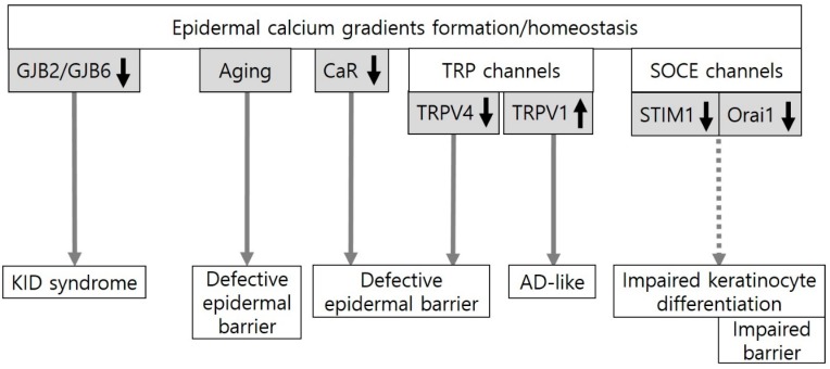
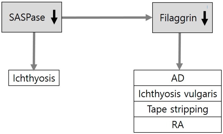
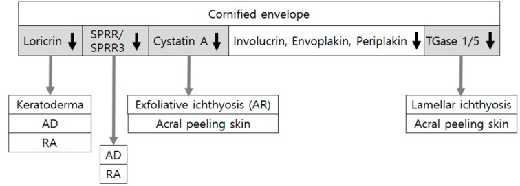
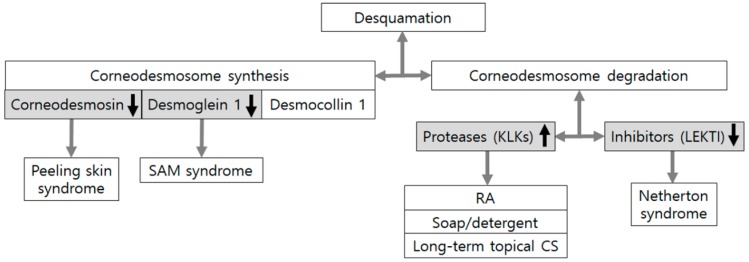
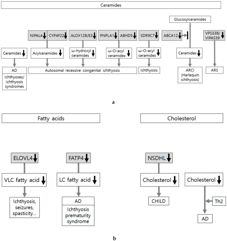
Epidermal barrier integrity could be influenced by various factors involved in epidermal cell differentiation and proliferation, cell-cell adhesion, and skin lipids. Dysfunction of this barrier can cause skin disorders, including eczema. Inversely, eczema can also damage the epidermal barrier. These interactions through vicious cycles make the mechanism complicated in connection with other mechanisms, particularly immunologic responses. In this article, the molecular mechanisms concerning epidermal barrier abnormalities are reviewed in terms of the following categories: epidermal calcium gradients, filaggrin, cornified envelopes, desquamation, and skin lipids. Mechanisms linked to ichthyoses, atopic dermatitis without exacerbation or lesion, and early time of experimental irritation were included. On the other hand, the mechanism associated with epidermal barrier abnormalities resulting from preceding skin disorders was excluded. The molecular mechanism involved in epidermal barrier dysfunction has been mostly episodic. Some mechanisms have been identified in cultured cells or animal models. Nonetheless, research into the relationship between the causative molecules has been gradually increasing. Further evidence-based systematic data of target molecules and their interactions would probably be helpful for a better understanding of the molecular mechanism underlying the dysfunction of the epidermal barrier.

Keywords: cornified envelopes; desquamation; epidermal calcium gradients; filaggrin; primary barrier dysfunction; skin lipids.


Conflict of interest statement

The author declare no conflict of interest.

Figures

Figure 1
Figure 1
An evidence-based or potential association between epidermal calcium gradients and the epidermal barrier. Epidermal calcium gradients have been altered in keratitis-ichthyosis-deafness (KID) syndrome induced by gap junction protein beta 2 (GJB2) or GJB6 missense mutations and in chronological skin aging. The formation and homeostasis of the epidermal calcium gradients could be regulated by the calcium-sensing receptor (CaR), transient receptor potential (TRP) channels, and store-operated calcium entry (SOCE) channels. CaR deletion, which inhibits Ca2+ influx, impairs the epidermal barrier in mice. In TRP channels, TRP vanilloid 4 (TRPV4) activation, which could be induced by heat (>30 °C) and hypo-osmolarity, plays an important role in epidermal barrier formation and recovery in mice. On the other hand, the blockade of TRPV1 activation by physical and chemical stimuli such as heat (42 °C) and capsaicin can suppress atopic dermatitis (AD)-like symptoms in mice. Two essential components of SOCE, STIM1 (stromal interaction molecule1) and Orai1 (ORAI calcium release-activated calcium modulator 1), are activated by endoplasmic reticulum (ER) calcium store depletion. Downregulation of Orai1 can impair keratinocyte differentiation and barrier homeostasis in mice.
Figure 2
Figure 2
Downregulation of filaggrin in skin diseases and experimental conditions related to epidermal barrier dysfunction. Loss-of-function mutations of FLG (encoding filaggrin) are associated with skin disorders related to barrier dysfunction, including atopic dermatitis (AD) and ichthyoses. Expression levels of filaggrin have also been reported to be reduced in experimental conditions of the disrupted epidermal barrier by tape stripping or retinoic acid (RA) application. SASPase (skin aspartic protease) generates filaggrin monomers from profilaggrin. Loss-of-function mutations of SASPase are associated with ichthyosis in dogs.
Figure 3
Figure 3
Downregulation of cornified envelope components in skin diseases and experimental conditions related to epidermal barrier dysfunction. Cornified envelopes are composed of various molecules, such as involucrin, loricrin, small proline-rich proteins (SPRRs), envoplakin, periplakin, and cysteine protease inhibitor A (cystatin A), crossed-linked by transglutaminases (TGases). Loss-of-function mutations in SPRR/SPRR3, LOR (encoding loricrin), TGM1/TGM5 (encoding transglutaminase1/5), and CSTA (encoding cystatin A) have been associated with atopic dermatitis (AD) or ichthyoses. Expression levels of loricrin and SPRRs have been reduced in experimental conditions of the disrupted epidermal barrier by retinoic acid (RA) application. Although triple knocking-out of envoplakin, periplakin, and involucrin induces abnormal cornified envelopes, mutations or deficiencies of involucrin alone do not cause atopic dermatitis.
Figure 4
Figure 4
Corneodesmosome defects in skin conditions associated with epidermal barrier dysfunction. Desquamation is determined by de novo synthesis and degradation of corneodesmosomal proteins. Mutations in GDSN encoding corneodesmosin and DSG1 encoding desmoglein 1 can cause an inflammatory type of peeling skin syndrome and SAM syndrome (severe skin dermatitis, multiple allergies, and metabolic wasting), which is another inflammatory type of peeling skin syndrome, respectively. Degradation of corneodesmosomal proteins depends on the sum of activities from proteases, including kallikrein-related peptidases (KLKs) and protease inhibitors including lymphoepithelial-Kazal-type 5 inhibitor (LEKTI). Experimental conditions, such as retinoic acid (RA) application, soap and detergent washing, and long-term corticosteroid (CS) application, could also accelerate desquamation mainly by increased production of KLKs.
Figure 5
Figure 5
Skin lipid alteration in skin conditions showing epidermal barrier dysfunction. (a) Skin lipids are composed of ceramides, free fatty acids, and cholesterol. Abnormalities in lipid composition, transport, and extracellular organization induce abnormal lipid organization. Most of the reports are related to abnormalities in ceramides, which are associated with ichthyoses, ichthyosis syndromes, and atopic dermatitis (AD). Loss-of-function mutations in ABCA12 (ATP-binding cassette transporter A12) cause harlequin ichthyosis, the most severe phenotype of autosomal recessive congenital ichthyosis (ARCI). Loss-of-function mutations in NIPAL4 (NIPA like domain containing 4), CYP4F22, ALOX12B and ALOXE3, PNPLA1 (Patatin-like phospholipase domain-containing lipase 1), and ABHD5 (α/β hydrolase domain-containing protein 5) also cause ARCI. (b) Not much has been reported about the association between altered synthesis of free fatty acid/cholesterol and skin barrier dysfunction. Loss-of-function mutations in ELOVL4 (fatty acid elongase 4) and FATP4 (fatty acid transport protein 4) reduce very long-chain (VLC) or LC fatty acids and cause neurocutaneous disorder (characterized by ichthyosis, seizures, spasticity, intellectual disability, and ichthyosis) and ichthyosis prematurity syndrome, respectively. Although loss-of-function mutations in NSDHL (NADP-dependent steroid dehydrogenase-like) inhibit cholesterol synthesis and cause CHILD syndrome (Congenital Hemidysplasia with Ichthyosiform Erythroderma and Limb Defects), cholesterol depletion is not considered as adequate to induce atopic dermatitis-like alteration in the absence of Th2 cytokines.

References

    1. Proksch E., Fölster-Holst R., Jensen J.M. Skin barrier function, epidermal proliferation and differentiation in eczema. J. Dermatol. Sci. 2006;43:159–169. doi: 10.1016/j.jdermsci.2006.06.003. - DOI - PubMed
    1. Chiang A., Tudela E., Maibach H.I. Percutaneous absorption in diseased skin: An overview. J. Appl. Toxicol. 2012;32:537–563. doi: 10.1002/jat.1773. - DOI - PubMed
    1. Kasemsarn P., Bosco J., Nixon R.L. The Role of the Skin Barrier in Occupational Skin Diseases. Curr. Probl. Dermatol. 2016;49:135–143. - PubMed
    1. Angelova-Fischer I. Irritants and Skin Barrier Function. Curr. Probl. Dermatol. 2016;49:80–89. - PubMed
    1. Fluhr J.W., Darlenski R., Angelova-Fischer I., Tsankov N., Basketter D. Skin irritation and sensitization: Mechanisms and new approaches for risk assessment. 1. Skin irritation. Skin Pharmacol. Physiol. 2008;21:124–135. doi: 10.1159/000131077. - DOI - PubMed