. 2020 Feb 11;9(2):151.
doi: 10.3390/antiox9020151.

The Exacerbation of Aging and Oxidative Stress in the Epididymis of Sod1 Null Mice

Affiliations

The Exacerbation of Aging and Oxidative Stress in the Epididymis of Sod1 Null Mice

Anaīs Noblanc et al. Antioxidants (Basel). .

Abstract

There is growing evidence that the quality of spermatozoa decreases with age and that children of older fathers have a higher incidence of birth defects and genetic mutations. The free radical theory of aging proposes that changes with aging are due to the accumulation of damage induced by exposure to excess reactive oxygen species. We showed previously that absence of the superoxide dismutase 1 (Sod1) antioxidant gene results in impaired mechanisms of repairing DNA damage in the testis in young Sod1-/- mice. In this study, we examined the effects of aging and the Sod-/- mutation on mice epididymal histology and the expression of markers of oxidative damage. We found that both oxidative nucleic acid damage (via 8-hydroxyguanosine) and lipid peroxidation (via 4-hydroxynonenal) increased with age and in Sod1-/- mice. These findings indicate that lack of SOD1 results in an exacerbation of the oxidative damage accumulation-related aging phenotype.

Keywords: 4-hydroxynonenal; 8-hydroxyguanosine; aging; epididymis; oxidative stress; reactive oxygen species; spermatozoa; superoxide dismutase.


Conflict of interest statement

None of the authors has any conflict of interest.

Figures

Figure 1
Figure 1
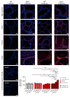
The histology of the epididymis is affected by aging in wild-type mice and even more so in Sod1−/− mice. Representative pictures of epididymal sections (initial segment, caput, corpus, and cauda epididymides) stained with toluidine blue from 3-month-old and 18-month-old wild-type and Sod1−/− mice. An increase in luminal round cells (circles) and in the number and size of clear vesicles (arrows) were observed in the epididymal epithelium of the corpus and cauda epididymides (n = 3). Scale bar: 40 µm.
Figure 2
Figure 2
Aging induces an accumulation the nucleic acid damage in the epididymis (initial segment, caput, corpus, and cauda epididymides) which is enhanced in Sod1−/− mice. Representative pictures of sections of the epididymis from 3-month-old and 18-month-old wild-type and Sod1−/− mice after immunostaining of oxidized nucleic acids (8-OHG, red); the nuclei are counterstained with DAPI (blue). Immunostaining negative controls (no primary antibody) are displayed for the caput and cauda epididymides. Scale bar: 40 µm. 8-OHG staining was quantified separately in the epididymal epithelium (clear histograms) and in the interstitial tissue (dashed histograms). ** p < 0.01, *** p < 0.001 (3-way ANOVA, n = 4–5).
Figure 3
Figure 3
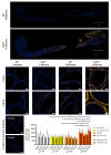
Lipid peroxidation as assessed by 4-HNE immunostaining is increased in the corpus and cauda epididymides with aging and this phenotype is enhanced in the absence of SOD1 expression. Representative pictures of whole epididymis sections of 18-month-old wild-type and Sod1−/− mice after the immunostaining of the 4-HNE (yellow) and the counterstaining of the nucleus (DAPI, blue). Scale bar: 2 mm. Observations have been carried out in epididymis sections of 3-month-old and 18-month-old wildtype and Sod1−/− mice. Staining was strongest in the corpus and cauda epididymides on the apical membrane of epithelial cells (arrow) and on intracellular membranes (arrow head). The negative controls (no primary antibody) for immunostaining are displayed for the corpus and cauda epididymides. Scale bar: 40 µm. The 4-HNE staining has been quantified in the epididymal epithelium (clear histograms) and in the interstitial tissue (dashed histograms) separately. * p < 0.05; ** p < 0.01 (3-way ANOVA, n = 4–5).

References

    1. Collin F. Chemical Basis of Reactive Oxygen Species Reactivity and Involvement in Neurodegenerative Diseases. Int. J. Mol. Sci. 2019;20:2407. doi: 10.3390/ijms20102407. - DOI - PMC - PubMed
    1. Ylä-Herttuala S. Oxidized LDL and Atherogenesis. Ann. New York Acad. Sci. 1999;874:134–137. doi: 10.1111/j.1749-6632.1999.tb09231.x. - DOI - PubMed
    1. Stadtman E.R., Levine R.L. Protein Oxidation. Ann. New York Acad. Sci. 2000;899:191–208. doi: 10.1111/j.1749-6632.2000.tb06187.x. - DOI - PubMed
    1. Marnett L.J. Oxyradicals and DNA Damage. Carcinogenesis. 2000;21:361–370. doi: 10.1093/carcin/21.3.361. - DOI - PubMed
    1. Wright W.W., Fiore C., Zirkin B.R. The Effect of Aging on the Seminiferous Epithelium of the Brown Norway Rat. J. Androl. 1993;14:110–117. - PubMed

LinkOut - more resources