. 2020 Jan-Dec;12(1):1725365.
doi: 10.1080/19420862.2020.1725365.

A functional antibody cross-reactive to both human and murine cytotoxic T-lymphocyte-associated protein 4 via binding to an N-glycosylation epitope

Affiliations

A functional antibody cross-reactive to both human and murine cytotoxic T-lymphocyte-associated protein 4 via binding to an N-glycosylation epitope

Dong Li et al. MAbs. 2020 Jan-Dec.

Abstract

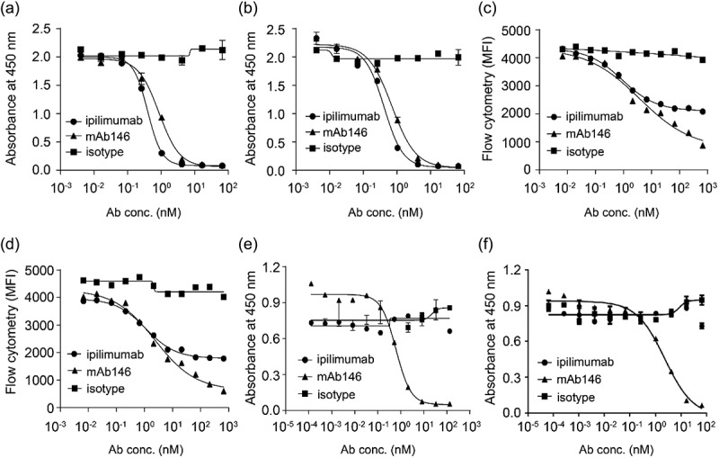
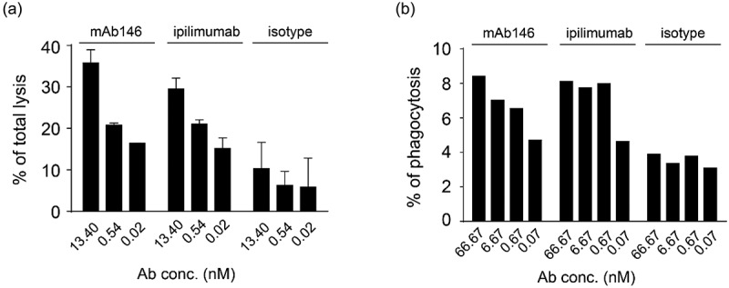
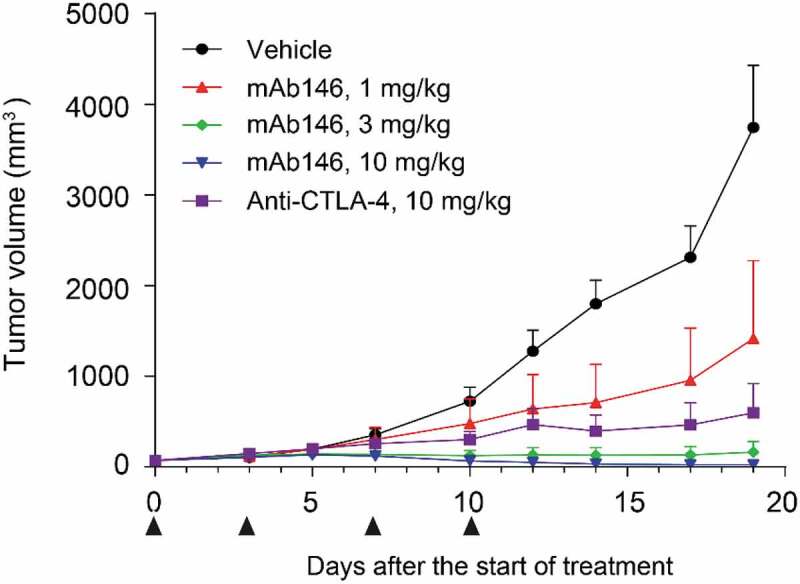
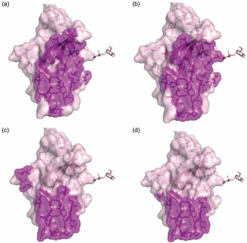
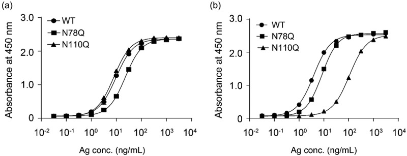
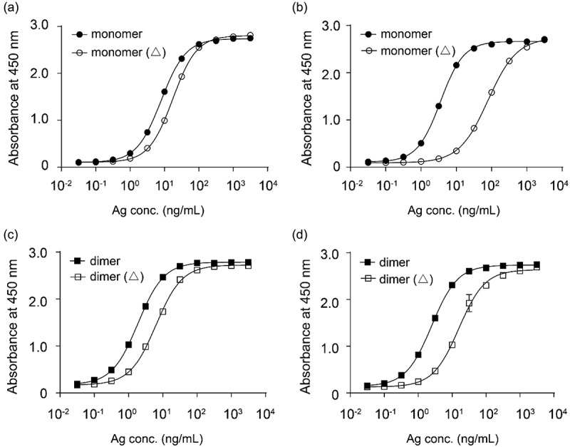
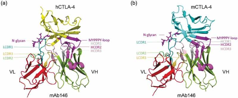
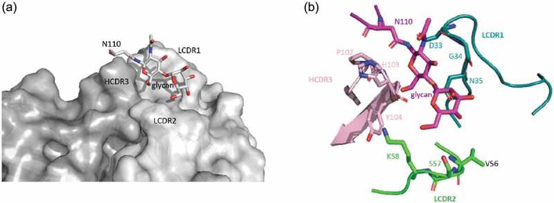
Cytotoxic T-lymphocyte-associated protein 4 (CTLA-4, CD152) is a receptor on T cells that inhibits the cell's functions. Blocking CTLA-4 with an antibody has proven effective for the treatment of cancer patients. Anti-CTLA-4 antibodies currently approved for clinical use can bind to human CTLA-4, but do not cross-react to murine CTLA-4. Here, we report the generation and characterization of a functional humanized antibody, mAb146, against both human and murine CTLA-4. Alanine scanning of CTLA-4 using mammalian cell expression cassette identified the unique epitopes of this novel antibody. In addition to the amino acid residues interacting with ligands CD80 and CD86, an N-glycosylation site on N110, conserved in CTLA-4 of human, monkey, and mouse, was identified as the specific epitope that might contribute to the cross-species binding and function of this antibody. This finding may also contribute to the understanding of the glycosylation of CTLA-4 and its related biologic function. In addition to facilitating preclinical development of anti-CTLA-4 antibodies, mAb146 may be useful as a therapeutic agent.

Keywords: CD152; Cytotoxic T-lymphocyte-associated protein 4 (CTLA-4); N-glycosylation; antibody; cross-species binding; epitope mapping.


Figures

Figure 1.
Figure 1.
Ipilimumab and mAb146 bound to human (a), monkey (b), and murine (c) CTLA-4 measured by ELISA. A 96-well plate was coated with hCTLA-4-6xHis monomer (1.0 μg/mL), cynomolgus monkey CTLA-4-6xHis monomer (0.5 μg/mL) or mouse CTLA-4-6xHis monomer (0.5 μg/mL) at 4C. After incubation with the antigens, the binding of Ipilimumab and mAb146 was detected by addition of HRP-conjugated goat anti-human IgG antibody.
Figure 2.
Figure 2.
Ipilimumab and mAb146 block ligands human CD80 (a, c) and human CD86 (b, d) binding to hCTLA-4 by ELISA (a–b) and FACS (c–d), and their ability on blocking mouse CD80 (e) and mouse CD86 (f) binding to mCTLA-4 also detected by ELISA (e–f). In ELISA-based competition assays, hCTLA-4-hFc dimer or mCTLA-4-mFc dimer (0.5 μg/mL) were coated on 96-well plates, the antibodies pre-mixed with 0.25 μg/mL of hCD80-6xHis, hCD86-6xHis, or 0.5 μg/ml of mCD80-6xHis, 5 μg/mL of mCD86-6xHis were added. After blocking with 2% BSA, biotinylated anti-His tag antibody was added. The bound ligands were detected by HRP-conjugated streptavidin. In FACS-based competition assay, hCD80- or hCD86-expressing CHO cell lines were used. The details were described in the method section.
Figure 3.
Figure 3.
Antibody-dependent cellular cytotoxicity (ADCC) (a) and antibody-dependent cellular phagocytosis (ADCP) (b) of the antibodies against CTLA-4. (a) Human CTLA4-expressing 293F cells were added to 96-well plates at 1 × 104 cells/well, and then the antibodies pre-incubated with 5 × 105 PBMCs were added. The plates were kept at 37°C in a 5% CO2 incubator for 4 h. Lysis of the target cells was determined by the introduction of DELFIA® EuTDA Cytotoxicity Reagents. (b) Human macrophage cells were mixed at 1:1 ratio with CFSE-dyed engineered human CTLA-4 expressing 293F cells in 96-well plates, then antibodies were added and incubated with cells at 37°C in a 5% CO2 incubator for 3 h. After wash, APC-labeled anti-human CD14 antibody was added for flow cytometry detection.
Figure 4.
Figure 4.
mAb146 significantly inhibited tumor growth in CT-26 syngeneic model. In CT-26 syngeneic model. Female Balb/C mice were inoculated subcutaneously with 1 × 105 tumor cells in 0.1 mL of PBS mixed with 50 μL matrigel. When the average tumor volume reached 60–80 mm3, the animals were randomly grouped (n = 6). The anti-CTLA-4 antibodies and isotype control were used for treatment. The tumor size was measured twice weekly by a vernier caliper, and tumor volume was calculated by the formula a× b2 × π/6 where “a” was length and “b” was width (a > b) of the tumor.
Figure 5.
Figure 5.
Hot spot residues or ligands binding sites mapped on CTLA-4 structure. Purple-shaded parts were identified as epitopes on human CTLA4. (a) Binding sites of Ipilimumab; (b) binding sites of antibody mAb146, the net indicates the N110 glycosylation site; (c) CD80 binding site (PDB code 1I8L); (d) CD86 binding site (PDB code 1I85).
Figure 6.
Figure 6.
Ipilimumab (a) and mAb146 (b) bound to wide type human CTLA-4 and glycosylation sites mutated (N78Q and N110Q) CTLA-4. The N-glycosylation sites N78 and N110 on CTLA-4 monomer with His tag were removed by single mutation of N78Q, N110Q. These CTLA-4 variants and WT CTLA-4 were incubated with 1 μg/ml of antibodies pre-coated on a 96-well plate. The bound CTLA4 variants were detected by HRP-labeled secondary antibody. The absorbance at 450 nM was measured using a microplate spectrophotometer.
Figure 7.
Figure 7.
Ipilimumab (a, c) and mAb146 (b, d) bound to monomeric human CTLA-4 with or without deglycosylation (a, b), and dimeric human CTLA-4 with or without deglycosylation (c, d). Each of the antibodies at concentration of 1 μg/ml was coated on 96-well plate overnight for ELISA binding assay. After interacting with untreated or PNase F-treated CTLA-4 protein, HRP-labeled secondary antibody was added as detection antibody. The absorbance at 450 nM was measured using a microplate spectrophotometer.
Figure 8.
Figure 8.
Structure model of the hCTLA-4:mAb146 and mCTLA-4:mAb 146 complexes. (a) The interface of hCTLA-4:mAb 146 complex is mainly composed of hCTHA-4 FG-loop (MYPPPY-loop, dark purple), N-glycan (dark purple) and mAb 146 CDRs. The FG loop mainly interacts with the HCDR2 (light purple). The HCDR1 (gray) mainly interacts with the C and C’ strands on hCTLA-4. The HCDR3 (pink), LCDR1 (cyan) and LCDR2 (light green) interact with the G strand, mainly the N-glycan. (b) The interface of mCTLA-4:mAb 146 is similar to that of hCTLA-4:mAb 146 complex. The color scheme is same as (A).
Figure 9.
Figure 9.
The binding of mAb146 with hCTLA-4 N110 glycan. (a) Surface representation of mAb 146 (heavy chain in dark gray; light chain in light gray) showing the pocket formed by HCDR3, LCDR1, and LCDR2. The N110 glycan inserts into the pocket. (b) Detailed interaction between the N110 glycan (purple) and mAb 146. P102, H103, Y104 on HCDR3 (pink), D33, G34, N35 on LCDR1 (cyan), and V56, S57, K58 on LCDR2 (green).
Figure 10.
Figure 10.
Structure comparison between human CTLA-4 and human CD28. The structures of hCTLA-4 (green) and hCD28 (salmon) are superposed. The main difference between these two is on the CC’-loop. On hCTLA-4, this loop interact with mAb146 HCDR1 and HCDR3, whereas a shorter loop on CD28 may cause potential clash. The L106 and I108 on hCTLA-4 and the corresponding residues D106 and E108 on CD28 are shown in sticks. The physical property of these residues may also contribute to the specificity of mAb146.

References

    1. Park J, Kwon M, Shin EC.. Immune checkpoint inhibitors for cancer treatment. Arch Pharm Res. 2016;39:1577–12. doi:10.1007/s12272-016-0850-5. - DOI - PubMed
    1. Qureshi OS, Zheng Y, Nakamura K, Attridge K, Manzotti C, Schmidt EM, Baker J, Jeffery LE, Kaur S, Briggs Z, et al. Trans-endocytosis of CD80 and CD86: a molecular basis for the cell-extrinsic function of CTLA-4. Science. 2011;332:600–03. doi:10.1126/science.1202947. - DOI - PMC - PubMed
    1. Rowshanravan B, Halliday N, Sansom DM. CTLA-4: a moving target in immunotherapy. Blood. 2018;131:58–67. doi:10.1182/blood-2017-06-741033. - DOI - PMC - PubMed
    1. Tang F, Du X, Liu M, Zheng P, Liu Y. Anti-CTLA-4 antibodies in cancer immunotherapy: selective depletion of intratumoral regulatory T cells or checkpoint blockade? Cell Biosci. 2018;8:30. doi:10.1186/s13578-018-0229-z. - DOI - PMC - PubMed
    1. Simpson TR, Li F, Montalvo-Ortiz W, Sepulveda MA, Bergerhoff K, Arce F, Roddie C, Henry JY, Yagita H, Wolchok JD, et al. Fc-dependent depletion of tumor-infiltrating regulatory T cells co-defines the efficacy of anti-CTLA-4 therapy against melanoma. J Exp Med. 2013;210:1695–710. doi:10.1084/jem.20130579. - DOI - PMC - PubMed

Publication types

LinkOut - more resources