. 2020 Feb 13;11(1):55.
doi: 10.1186/s13287-020-1567-4.

Multipotency of mouse trophoblast stem cells

Affiliations

Multipotency of mouse trophoblast stem cells

Minmin Hou et al. Stem Cell Res Ther. .

Abstract

Background: In a number of disease processes, the body is unable to repair injured tissue, promoting the need to develop strategies for tissue repair and regeneration, including the use of cellular therapeutics. Trophoblast stem cells (TSCs) are considered putative stem cells as they differentiate into other subtypes of trophoblast cells. To identify cells for future therapeutic strategies, we investigated whether TSCs have properties of stem/progenitor cells including self-renewal and the capacity to differentiate into parenchymal cells of fetal organs, in vitro and in vivo.

Methods: TSCs were isolated using anti-CD117 micro-beads, from embryonic day 18.5 placentas. In vitro, CD117+ TSCs were cultured, at a limiting dilution in growth medium for the development of multicellular clones and in specialized medium for differentiation into lung epithelial cells, cardiomyocytes, and retinal photoreceptor cells. CD117+ TSCs were also injected in utero into lung, heart, and the sub-retinal space of embryonic day 13.5 fetuses, and the organs were harvested for histological assessment after a natural delivery.

Results: We first identified CD117+ cells within the labyrinth zone and chorionic basal plate of murine placentas in late pregnancy, embryonic day 18.5. CD117+ TSCs formed multicellular clones that remained positive for CD117 in vitro, consistent with self-renewal properties. The clonal cells demonstrated multipotency, capable of differentiating into lung epithelial cells (endoderm), cardiomyocytes (mesoderm), and retinal photoreceptor cells (ectoderm). Finally, injection of CD117+ TSCs in utero into lungs, hearts, and the sub-retinal spaces of fetuses resulted in their engraftment on day 1 after birth, and the CD117+ TSCs differentiated into lung alveolar epithelial cells, heart cardiomyocytes, and retina photoreceptor cells, corresponding with the organs in which they were injected.

Conclusions: Our findings demonstrate that CD117+ TSCs have the properties of stem cells including clonogenicity, self-renewal, and multipotency. In utero administration of CD117+ TSCs engraft and differentiate into resident cells of the lung, heart, and retina during mouse development.

Keywords: In utero; Multipotency; Stem cells; Trophoblast cells.


Conflict of interest statement

The authors declare that they have no competing interests.

Figures

Fig. 1
Fig. 1
Expression of CD117 in trophoblast cells in labyrinth zone and basal chorionic plate of placenta. a Representative image of nuclear staining for hematoxylin, showing anatomical structure of an embryonic (E) day 18.5 placenta (top panel). A representative image of immunofluorescence staining for CD117 (green) and DAPI (nuclei, blue) from labyrinth region (second panel) depicted in the green box of the top panel. A representative image of immunohistochemical staining for CD117 (brown) and hematoxylin for nuclei (third panel), from the chorion depicted in the brown box in the top image. Bottom panels are representative images of immunofluorescence staining for CD117 (green, left) and CK7 (red, middle) and merged image with for nuclei. b Representative image of immunofluorescence co-staining for CD117 (green, left) with Ki67 (red, middle), merged image with DAPI (right). c Representative immunofluorescence image of co-staining for CD117 (green, left column) and markers for hematopoietic cells, CD34, CD45, and CD31 (red, middle column, respectively), and merged with DAPI (right column). d Representative immunofluorescence image of co-staining of CD117 (green, left column) and markers for MSCs, vimentin, and E-cadherin (VMT and ECAM, respectively, red, middle column) and merged with DAPI (right column)
Fig. 2
Fig. 2
Characterization of CD117+ TSCs in vitro. Clonal CD117+ TSCs were harvested for flow cytometric analysis. a Representative scatter plots of flow cytometer for CD90 versus Sca1 (first panel), CD105 versus CD73 (second panel), CD31 versus CD34 (third panel), and CD11b versus CD45 (forth panel). b Quantitation of the flow cytometric assay showing percentage of markers for MSCs (open bars), Sca1, CD90.2, CD 105, and CD73 and hematopoietic cells (black bars), CD34, CD31, CD11b, and CD45, in total placental cells, n = 3~4 for each marker. c Representative histogram of flow cytometric assays for major histocompatibility complex classic I and II (MHC I and MHC II, respectively), gray for APC isotope control, red for MHC I and II staining (left and right, respectively)
Fig. 3
Fig. 3
Self-renewal of placental CD117+ TSCs in vitro. CD117+ TSCs were plated in a 100-mm dish at limited dilution of one cell every 60 mm2 for 14 days. a Representative image of multicellular clones stained for crystal violet (left, purple), higher magnification of a clone staining for crystal violet (middle, purple), and phase-contrast image of an entire clone (right). b Representative image of a clone staining positive for CD117 (green), DAPI for nuclear staining. c After expansion, 97% of clonal cells are positive for CD117 as represented in flow cytometry histogram, gray for Alex 488 isotope control, red for CD117 Alex 488 staining. d Representative image of clonal cells staining positive for caudal type homeobox 2 (CDX2; green, left), DAPI for nuclear staining (blue, middle), and merged image (right)
Fig. 4
Fig. 4
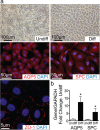
Differentiation of CD117+ TSCs into alveolar epithelial cells of lung in vitro. a Phase-contrast images showing morphologic change of cells before (Undiff, top row, left) and after differentiation (Diff, top row, right) in epithelial cell differentiation medium on air-liquid interface for 14 days. These differentiated cells were immunostained for AQP5 (red, middle row, left), SPC (red, middle row, right), and ZO-1 (red, bottom row, left). b Bar graph showing the quantitation of qRT-PCR for AQP5 and SPC in fold change comparing differentiated (Diff, black bars) to undifferentiated cells (Undiff, open bar). *P < 0.05 compared to their undifferentiated cells, n = 5 for each group
Fig. 5
Fig. 5
Differentiation of CD117+ TSCs into cardiomyocytes in vitro.a Phase-contrast images showing morphologic change of cells before (Undiff, top row, left) and after differentiation (Diff, top row, right) in cardiomyocyte differentiation medium for 12 days. After differentiation, representative images of immunofluorescence staining for cTnT (green, middle row, left) and Sar α-actinin (green, middle row, right), merged with DAPI for nuclear staining (blue). Bottom panels show the confocal images of differentiated cells staining for cTnT (white, bottom row, left) and Sar α-actinin (white, bottom row, right). Red arrows highlight areas of striations. b Bar graph showing the quantitation of qRT-PCR for cTnT and Sar α-actinin in fold change comparing differentiated (Diff, black bars) to undifferentiated cells (Undiff, open bar). * P < 0.05 compared to their undifferentiated cells, n = 4~5 for each group
Fig. 6
Fig. 6
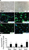
Differentiation of CD117+ TSCs into retinal photoreceptor cells in vitro. a Phase-contrast images showing morphologic change of cells before (Undiff, top row, left) and after differentiation (Diff, top row, right) in neuronal differentiation medium for 8 days. After differentiation, representative image of immunofluorescence staining of differentiated cells for Rho (green, middle row, left), Rec (green, middle row, right), and merged with DAPI for nuclear staining (blue). Arrestin 1 (Arr1, green, bottom row, left) and merged with DAPI for nuclear staining (blue, bottom row, right). b Bar graph showing quantitation of qRT-PCR for Rho, Rec, Arr 1 and Arrestin 4 (Arr4) comparing fold change in differentiated (Diff, black bars) versus undifferentiated cells (Undiff, open bar). *P < 0.05 compared to their undifferentiated cells, n = 4 for each group
Fig. 7
Fig. 7
Differentiation of CD117+ TSCs into three germ layers in vitro. CD117+ TSCs were cultured by a hanging drop technique for 2 days and then the spheroids were cultured in ultra-low attachment 6-well plates for 14 days in growth medium in the absence of LIF and FGFβ. Total RNA was then extracted from the cells. a Bar graph showing quantitation of qRT-PCR for fold change in markers of endoderm (CER1 and GATA6), mesoderm (TBXT and BMP7), and ectoderm (FGF5 and OTX2). b Bar graph showing quantitation of qRT-PCR for fold change in markers of endoderm-derived lung alveoli epithelium cells (AQP5 and SPC), mesoderm-derived cardiomyocytes cells (Sar α-actinin and cTnT), and ectoderm-derived retinal photoreceptor cells (Rho and Rec). *P < 0.05 compared to their undifferentiated cells, n = 4 for each group
Fig. 8
Fig. 8
Differentiation of CD117+ TSCs into three germ layers in vivo. Under ultrasound guidance (left column), CD117+ TSCs pre-labeled with PKH67 (green) were injected in utero into lung, myocardium, or the sub-retinal space of E13.5 mouse fetuses. Lungs, hearts, and eyes were harvested at day 1 after nature birth for histological assessment. Representative merged images of exogenous cells (TSCs, green), DAPI (blue), and immunofluorescence staining for a marker of lung alveolar epithelium, SPC (red, upper row), or a marker of cardiomyocytes, cTnI (red, middle row), or a marker of retinal photoreceptors, Rho (red, lower row). The third and fourth columns show respectively higher power magnification images of the area in the white boxes of the second column

References

    1. Trounson A, McDonald C. Stem cell therapies in clinical trials: progress and challenges. Cell Stem Cell. 2015;17:11–22. doi: 10.1016/j.stem.2015.06.007. - DOI - PubMed
    1. Trounson A, Thakar RG, Lomax G, Gibbons D. Clinical trials for stem cell therapies. BMC Med. 2011;9:52. doi: 10.1186/1741-7015-9-52. - DOI - PMC - PubMed
    1. Brooke G, et al. Manufacturing of human placenta-derived mesenchymal stem cells for clinical trials. Br J Haematol. 2009;144:571–579. doi: 10.1111/j.1365-2141.2008.07492.x. - DOI - PubMed
    1. Lublin FD, et al. Human placenta-derived cells (PDA-001) for the treatment of adults with multiple sclerosis: a randomized, placebo-controlled, multiple-dose study. Mult Scler Relat Disord. 2014;3:696–704. doi: 10.1016/j.msard.2014.08.002. - DOI - PubMed
    1. Phan TG, Ma H, Lim R, Sobey CG, Wallace EM. Phase 1 trial of amnion cell therapy for ischemic stroke. Front Neurol. 2018;9:198. doi: 10.3389/fneur.2018.00198. - DOI - PMC - PubMed

Publication types