. 2020 Feb 13;10(1):2521.
doi: 10.1038/s41598-020-59313-8.

The anti-tumoral potential of the saporin-based uPAR-targeting chimera ATF-SAP

Affiliations

The anti-tumoral potential of the saporin-based uPAR-targeting chimera ATF-SAP

S Zuppone et al. Sci Rep. .

Abstract

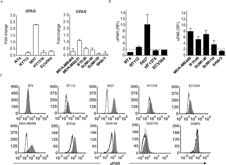
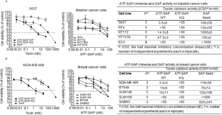
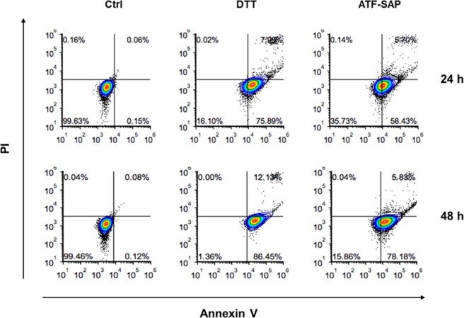
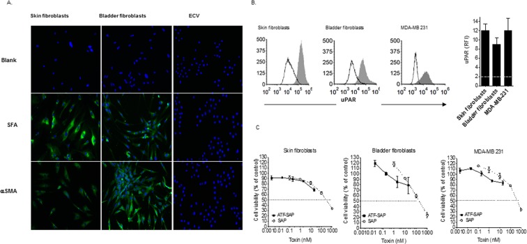
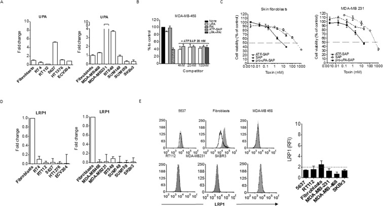
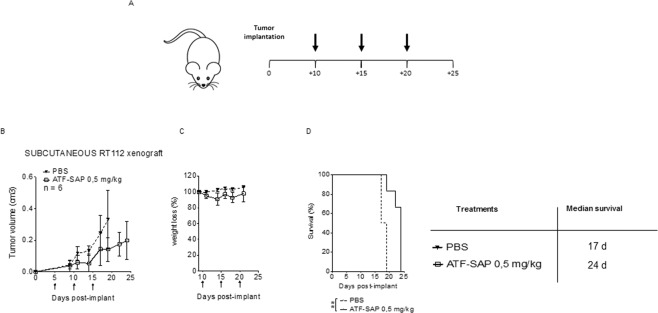
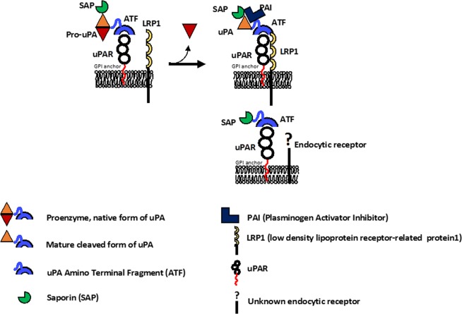
The development of personalized therapies represents an urgent need owing to the high rate of cancer recurrence and systemic toxicity of conventional drugs. So far, targeted toxins have shown promising results as potential therapeutic compounds. Specifically, toxins conjugated to antibodies or fused to growth factors/enzymes have been largely demonstrated to selectively address and kill cancer cells. We investigated the anti-tumor potential of a chimeric recombinant fusion protein formed by the Ribosome Inactivating Protein saporin (SAP) and the amino-terminal fragment (ATF) of the urokinase-type plasminogen activator (uPA), whose receptor has been shown to be over-expressed on the surface of aggressive tumors. ATF-SAP was recombinantly produced by the P. pastoris yeast and its activity was assessed on a panel of bladder and breast cancer cell lines. ATF-SAP resulted to be highly active in vitro, as nano-molar concentrations were sufficient to impair viability on tumor cell lines. In contrast to untargeted toxins, the chimeric fusion protein displayed a significantly improved toxic effect in uPAR-expressing cells, demonstrating that the selective activity was due to the presence of the targeting moiety. Fibroblasts were not sensitive to ATF-SAP despite uPAR expression, indicating that cell-specific receptor-mediated internalization pathway(s) might be considered. The in vivo anti-tumor effect of the chimera was shown in a bladder cancer xenograft model. Current findings indicate ATF-SAP as a suitable anti-tumoral therapeutic option to cope with cancer aggressiveness, as a single treatment or in combination with traditional therapeutic approaches, to appropriately address the intra- and inter- tumor heterogeneity.


Conflict of interest statement

The authors declare no competing interests.

Figures

Figure 1
Figure 1
UPAR expression in bladder and breast cancer cell lines. Bladder and breast cancer cell lines resembling respectively different grades (RT4 -grade 1-, RT112 and 5637- grade 2-, HT1376 and ECV304 -grade 3-) and subtypes (MDA-MB-468, BT549, SUM149, SUM159 -TNBC-, SKBR3 -HER2+-) were analyzed by quantitative PCR (see “Materials and Methods”) for UPAR gene expression (A) and by flow cytometry for uPAR protein expression (b and c). UPAR protein expression is shown as histogram plots (B) and Relative Fluorescence Intensity (RFI) (see “Materials and Methods”) (C). SKBR3 HER2+ breast cancer cell line was used as subtype control. The dashed line represents the threshold arbitrarily defining positive expression (RFI = 2).
Figure 2
Figure 2
Cytotoxic activity of ATF-SAP on bladder and breast cancer cells. ATF-SAP activity and target specificity were evaluated on RT4, RT112, 5637, HT1376 and ECV304 bladder cancer cell lines (A) and on MDA-MB-468, SUM149, SUM159, BT549 TNBC and HER2+ SKBR3 breast cancer cell lines. (B) Cells were incubated for 72 h with scalar logarithmic concentrations of the toxin and cell viability was analyzed by MTT assay. The untargeted seed SAP and the catalytically inactive mutant ATF-SAP KQ were used as controls. The IC50 from three different experiments is reported as mean ± SE.
Figure 3
Figure 3
ATF-SAP cell apoptosis induction. 5637 bladder cancer cells were incubated with ATF-SAP for 24 or 48 hours. Flow cytometry analysis was performed to distinguish early apoptotic (lower right gate) form late apoptotic (upper right) and necrotic (upper left) cell populations.
Figure 4
Figure 4
ATF-SAP activity on fibroblasts and MDA-MB-231 cancer cell line. (A) Molecular profile of skin and bladder-derived fibroblasts. Human skin- and bladder-derived primary fibroblasts were analyzed by immunofluorescence for the expression of Fibroblasts Surface Antigen (SFA) and alpha-Smooth Muscle Actin (αSMA). ECV304 epithelial bladder cancer cell line was used as negative control. Nuclei were stained with DAPI (blue). (B) uPAR expression on skin and bladder-derived fibroblasts as well as breast cancer MDA-MB-231 cell line was analyzed by flow cytometry and expressed as histogram plots (upper panel) and Relative Fluorescence Intensity (RFI) (see “Materials and Methods”) (lower panel). The dashed line represents the threshold arbitrarily defining positive expression (RFI = 2). (C) ATF-SAP toxic activity was evaluated after 72 h incubation and compared to the untargeted seed SAP. Results from one representative experiment are shown as mean ± SD. Three independent experiments were performed for each assay.
Figure 5
Figure 5
UPA and LRP1 expression in bladder and breast cancer cell lines. (A) Skin fibroblasts, bladder and breast cancer cell lines respectively resembling different stages (RT4 -T1 superficial-, RT112 and 5637- T2 muscle invasive-, HT1376 and ECV304 -T3 muscle invasive-) and subtypes (MDA-MB-468, MDA-MB-231, BT549, SUM149, SUM159 -TNBC-, SKBR3 -HER2+-) were analyzed by qPCR for UPA gene expression (see “Materials and Methods”). (B) Competition assay between ATF-SAP and uPAR natural ligands. MDA-MB-468 cells were incubated with ATF-SAP 20 nM in the presence of equal or increasing concentrations of uPA or PAI. The effect on cell viability was evaluated after 72 h by MTT assay. Seed SAP was used as untargeted control. (C) Comparison of pro-uPA-SAP and ATF-SAP toxic activity on fibroblasts and MDA-MB-231 cells. Results from one representative experiment are shown as mean ± SD. Three independent experiments were performed for each assay. LRP1 gene (D) and protein (E) expression on bladder and breast cancer cells. LRP1 protein expression is displayed as histogram plots (E, left panel) and RFI (see “Materials and Methods”) (E, right panel). The dashed line represents the threshold arbitrarily defining positive expression (RFI = 2).
Figure 6
Figure 6
Cytotoxic activity of ATF-SAP on ex vivo bladder cancer cells. (A) After the second implantation, established tumors were minced and tumor cells analyzed for uPAR expression by flow cytometry. Results are shown as RFI (see “Materials and Methods”) (left panel). The dashed line represents the threshold arbitrarily defining positive expression (RFI = 2). (B) Explanted cells were then incubated for 72 h with ATF-SAP and cell viability was evaluated by MTT assay (right panels). Seed SAP was used as untargeted control. Results from one representative experiment are shown as mean ± SD. Three independent experiments were performed.
Figure 7
Figure 7
Effects of ATF-SAP on tumor growth in a subcutaneous xenograft bladder cancer mouse model. (A) Schematic representation of the experimental design. RT112 bladder cancer cells were subcutaneously injected in 7-week-old nude female mice. Tumor bearing mice were randomized into 2 experimental groups and treated respectively with PBS or ATF-SAP (0.5 mg/Kg) via intravenous injection every 5 days after tumor implantation. (B) Quantitative analysis of growing tumor volume in mice are shown as mean ± SD from respectively n = 6 mice per condition. (C) The mean weight of mice from each treatment group is shown as percentage from initial. (D) Kaplan–Meyer plot of animal survival with median survival time listed in the table. Results from a Mantel–Cox two-sided log-rank test are shown when statistically significant (**p < 0.01) for ATF-SAP 0.5 mg/kg (red; hazard ratio, 19.2; 95% CI, 2.2–165.8) versus PBS in mice bearing subcutaneous RT112 tumor.
Figure 8
Figure 8
Schematic representation of internalization pathways proposed for uPAR targeting toxins. (A) LRP1-uPA-PAI dependent endocytosis; (B) LRP1-uPA-PAI independent endocytosis.

References

    1. Gatzka Martina. Targeted Tumor Therapy Remixed—An Update on the Use of Small-Molecule Drugs in Combination Therapies. Cancers. 2018;10(6):155. doi: 10.3390/cancers10060155. - DOI - PMC - PubMed
    1. Holohan C, Van Schaeybroeck S, Longley DB, Johnston PG. Cancer drug resistance: an evolving paradigm. Nat Rev Cancer. 2013;13:714–726. doi: 10.1038/nrc3599. - DOI - PubMed
    1. Vago, R., Ippoliti, R., Fabbrini, M. S. In Antitumor Potential and other Emerging Medicinal Properties of Natural Compounds (ed Evandro Fei Fang; Tzi Bun Ng) 145–179 (Springer, 2013).
    1. Fabbrini Maria, Katayama Miku, Nakase Ikuhiko, Vago Riccardo. Plant Ribosome-Inactivating Proteins: Progesses, Challenges and Biotechnological Applications (and a Few Digressions) Toxins. 2017;9(10):314. doi: 10.3390/toxins9100314. - DOI - PMC - PubMed
    1. Cavallaro U, Nykjaer A, Nielsen M, Soria MR. Alpha 2-macroglobulin receptor mediates binding and cytotoxicity of plant ribosome-inactivating proteins. Eur J Biochem. 1995;232:165–171. doi: 10.1111/j.1432-1033.1995.tb20795.x. - DOI - PubMed

Publication types

MeSH terms

Substances