. 2020 Jan 7:18:199-206.
doi: 10.1016/j.csbj.2019.12.012. eCollection 2020.

The Glu102 mutation disrupts higher-order oligomerization of the sigma 1 receptor

Affiliations

The Glu102 mutation disrupts higher-order oligomerization of the sigma 1 receptor

Ara M Abramyan et al. Comput Struct Biotechnol J. .

Abstract

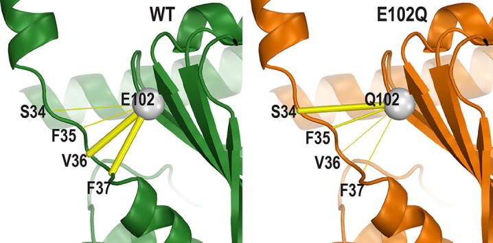
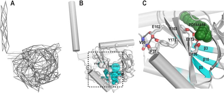
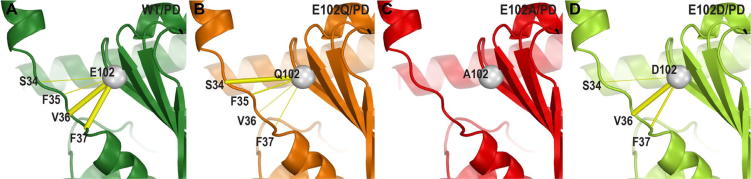
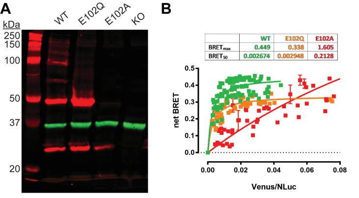
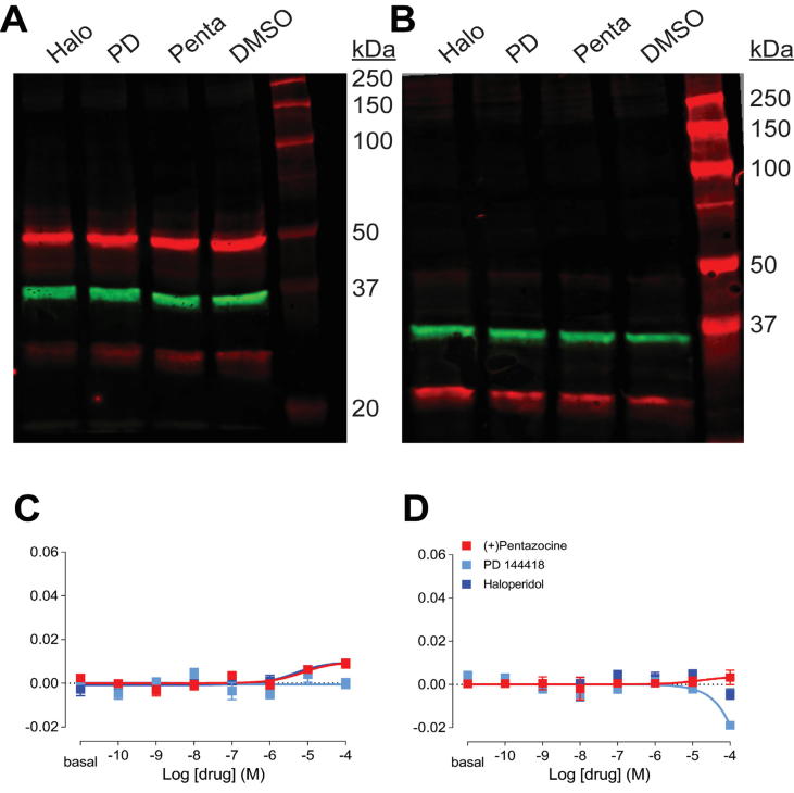
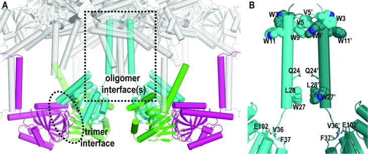
The sigma 1 receptor (σ1R) is a unique endoplasmic reticulum membrane protein. Its ligands have been shown to possess therapeutic potential for neurological and substance use disorders among others. The E102Q mutation of σ1R has been found to elicit familial cases of amyotrophic lateral sclerosis (ALS). Despite reports of its downstream signaling consequences, the mechanistic details of the functional impact of E102Q at molecular level are not clear. Here, we investigate the molecular mechanism of the E102Q mutation with a spectrum of biochemical, biophysical, and pharmacological approaches. Our analysis of the interaction network of σ1R indicates that a set of residues near E102 is critical for the integrity of C-terminal ligand-binding domain. However, this integrity is not affected by the E102Q and E102A mutations, which is confirmed by the radioligand binding results. Instead, the E102 mutations disrupt the connection between the C-terminal domain and the N-terminal transmembrane helix (NT-helix). Results from bioluminescence resonance energy transfer and western blot assays demonstrate that these mutations destabilize higher-order σ1R oligomers, while our molecular dynamics simulations based on a σ1R crystal structure reveal a potential mechanism by which the mutations perturb the NT-helix dynamics. Thus, we propose that E102 is at a critical position in propagating the effects of ligand binding from the C-terminal domain to the NT-helix, while the latter may be involved in forming alternative oligomer interfaces, separate from the previously reported trimer interface. Together, these results provide the first account of the molecular mechanism of σ1R dysfunction caused by E102Q.

Keywords: Amyotrophic lateral sclerosis; Bioluminescence resonance energy transfer; Molecular dynamics simulations; Oligomerization; Sigma 1 receptor; Western blot.


Conflict of interest statement

The authors declare that they have no known competing financial interests or personal relationships that could have appeared to influence the work reported in this paper.

Figures

None
Graphical abstract
Fig. 1
Fig. 1
E102 is closely associated with the structural motif connecting the NT-helix and the C-terminal domain. (A) Contacts in the σ1R WT/PD144418 crystal structure (PDB ID 5HK1) are represented as lines connecting the contacting residue pairs. (B) The eigenvector centrality score of each residue (see Materials and Methods) in the σ1R WT/PD144418 crystal structure C-terminal domain is represented by the radius of the sphere. The residues with the highest centrality scores (Fig. S1B) are colored in cyan. (C) A zoom-in view of the ligand binding pocket showing the interactions between the sidechain of E102 and the loop connecting the N-terminal transmembrane helix with the C-terminal domain. The nearby residues essential to ligand binding are shown as sticks as well. PD144418 is represented in green spheres. The positions of the high-centrality residues shown in panel B are colored on the backbone ribbons. (For interpretation of the references to colour in this figure legend, the reader is referred to the web version of this article.)
Fig. 2
Fig. 2
The E102 mutations disrupt the connection between the NT-helix and the C-terminal domain. The Cα atom of residue 102 is shown as a gray sphere and its contacts are shown in yellow sticks with varied thickness proportional to their frequency of the polar interactions to the loop region (residues 34–37) in the simulated conditions. Protein backbone ribbons are colored in green, orange, red, and light green for WT/PD144418 (PD) (A), E102Q/PD (B), E102A/PD (C), and E102D/PD (D) conditions, respectively. (For interpretation of the references to colour in this figure legend, the reader is referred to the web version of this article.)
Fig. 3
Fig. 3
WT σ1R forms high-order oligomers while E102Q and E102A do not. (A) Representative western blot from five experiments are shown to visualize σ1R (red) and GAPDH (green) bands for wildtype (WT), E102Q, and E102A constructs expressed in Δσ1R HEK293T cells. Protein extract from Δσ1R cells without σ1R rescue expression is run in the last lane. (B) Molecular interactions between σ1R monomers are measured by BRET. Δσ1R HEK 293T cells were transfected with a constant amount of the RLuc-fusion construct and increasing amounts of the Venus-fusion construct (green-WT, orange-E102Q, and red-E102A). All data points were performed in triplicate (S.E.M. shown as error bars). The BRETmax and BRET50 values were calculated by nonlinear regression using a single-site saturation binding model. (For interpretation of the references to colour in this figure legend, the reader is referred to the web version of this article.)
Fig. 4
Fig. 4
E102 mutations abrogated drug response in σ1R oligomerization. (A-B) Representative western blots from five experiments are shown to visualize σ1R (red) and GAPDH (green) bands after drug treatment for E102Q (A) and E102A (B) constructs expressed in Δσ1R HEK293T cells. (C-D) Drug-induced changes in σ1R homomer BRET ratios are shown for the E102Q (C) and E102A (D) constructs expressed in Δσ1R HEK293T cells. None of (+)-pentazocine (red), PD144418 (cyan), or haloperidol (blue) induced significant changes in either of the mutants. Data represent mean ± S.E.M. (n = 5 or more). (For interpretation of the references to colour in this figure legend, the reader is referred to the web version of this article.)
Fig. 5
Fig. 5
The crystal structures of σ1R may include other oligomer interface(s). (A) In addition to the trimer interface reported previously , analysis of the asymmetric units in a representative crystal structure of σ1R (PDB ID 5HK1) shows that the NT-helices from 6 monomers cluster together (in the dotted box), with two of them (cyan) in inverted orientation from the other four (gray). Although overall this cluster of NT-helices is an artifact of crystallography, it may include other oligomeric interface(s) that are potentially functionally relevant, such as that in panel B. The backbone ribbons of three monomers in a trimer are colored in green, cyan, and magenta. (B) In this symmetric dimer interface (dotted box in panel A), three Trp and one Val residues from each monomer are tightly packed together at their N-termini, while several other residues face each other within an interacting range at the bottom of these two NT-helices. E102 interacts with the backbone amine groups in the loop connecting the NT-helix and C-terminal domain. (For interpretation of the references to colour in this figure legend, the reader is referred to the web version of this article.)

References

    1. Hayashi T., Su T.P. Sigma-1 receptor chaperones at the ER-mitochondrion interface regulate Ca(2+) signaling and cell survival. Cell. 2007;131(3):596–610. - PubMed
    1. Su T.P. The Sigma-1 receptor as a pluripotent modulator in living systems. Trends Pharmacol Sci. 2016;37(4):262–278. - PMC - PubMed
    1. Stone J.M. [123I]TPCNE–a novel SPET tracer for the sigma-1 receptor: first human studies and in vivo haloperidol challenge. Synapse. 2006;60(2):109–117. - PubMed
    1. Katz J.L. A role for sigma receptors in stimulant self-administration and addiction. Behav Pharmacol. 2016;27(2-3 Spec Issue):100–115. - PMC - PubMed
    1. Hashimoto K. Sigma-1 receptor chaperone and brain-derived neurotrophic factor: emerging links between cardiovascular disease and depression. Prog Neurobiol. 2013;100:15–29. - PubMed