

Effective dose and image optimisation of lateral lumbar spine radiography: a phantom study
- PMID: 32056045
- PMCID: PMC7018898
- DOI: 10.1186/s41747-019-0132-3
Effective dose and image optimisation of lateral lumbar spine radiography: a phantom study
Abstract
Background: To investigate lateral lumbar spine radiography technical parameters for reduction of effective dose whilst maintaining image quality (IQ).
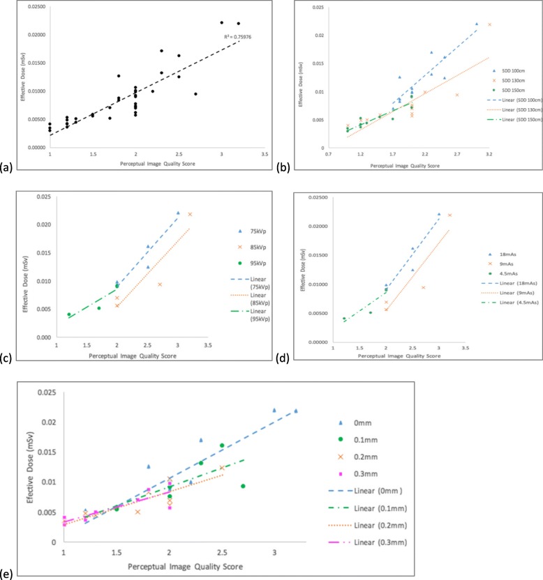
Methods: Thirty-six radiograms of an anthropomorphic phantom were acquired using different exposure parameters: source-to-detector distance (SDD) (100, 130 or 150 cm), tube potential (75, 85 or 95 kVp), tube current × exposure time product (4.5, 9, 18 mAs) and additional copper (Cu) filter (no filter, 0.1-, 0.2-, or 0.3-mm thickness. IQ was assessed using an objective approach (contrast-to-noise-ratio [CNR] calculation and magnification measurement) and a perceptual approach (six observers); ED was estimated using the PCXMC 2.0 software. Descriptive statistics, paired t test, and intraclass correlation coefficient (ICC) were used.
Results: The highest ED (0.022 mSv) was found with 100 cm SSD, 75 kVp, 18 mAs, and without Cu filter, whilst the highest CNR (7.23) was achieved at 130 cm SSD, 75 kVp, 18 mAs, and without Cu filter. The lowest ED and CNR were generated at 150 cm SDD, 95 kVp, 4.5 mAs, and 0.3-mm Cu filter. All observers identified the relevant anatomical structures on all images with the lowest ED and IQ. The intra-observer (0.61-0.79) and inter-observer (0.55-0.82) ICC ranged from moderate to excellent.
Conclusion: All relevant anatomical structures were identified on the lateral lumbar spine radiographs despite using low-dose protocols. The lowest ED (0.002 mSv) was obtained with 150 cm SDD, 95 kVp, 4.5 mAs, and 0.3-mm Cu filter. Further technical and clinical studies are needed to verify these preliminary findings.
Keywords: Image quality; Lumbosacral region; Phantoms (imaging); Radiation dosage; Radiography.
Conflict of interest statement
The authors declare that they have no competing interests.
Figures

References
-
- American College of Radiology (2017) Practice parameter for the performance of spine radiography. Available via https://www.acr.org/-/media/ACR/Files/Practice-Parameters/rad-spine.pdf?...
-
- Davey Enda, England Andrew. AP versus PA positioning in lumbar spine computed radiography: Image quality and individual organ doses. Radiography. 2015;21(2):188–196. doi: 10.1016/j.radi.2014.11.003. - DOI
-
- New South Wales (NSW) Agency for Clinical Innovation (2012) Radiology Clinician Fact Sheet. Available via https://www.aci.health.nsw.gov.au/__data/assets/pdf_file/0006/174552/MI-.... Accessed 16 Oct 2018
MeSH terms
LinkOut - more resources
Full Text Sources