Review
. 2020 Jun;26(3):249-271.
doi: 10.1089/ten.teb.2019.0302. Epub 2020 Feb 14.

Modeling the Tumor Microenvironment and Pathogenic Signaling in Bone Sarcoma

Affiliations
Review

Modeling the Tumor Microenvironment and Pathogenic Signaling in Bone Sarcoma

Eric R Molina et al. Tissue Eng Part B Rev. 2020 Jun.

Abstract

Investigations of cancer biology and screening of potential therapeutics for efficacy and safety begin in the preclinical laboratory setting. A staple of most basic research in cancer involves the use of tissue culture plates, on which immortalized cell lines are grown in monolayers. However, this practice has been in use for over six decades and does not account for vital elements of the tumor microenvironment that are thought to aid in initiation, propagation, and ultimately, metastasis of cancer. Furthermore, information gleaned from these techniques does not always translate to animal models or, more crucially, clinical trials in cancer patients. Osteosarcoma (OS) and Ewing sarcoma (ES) are the most common primary tumors of bone, but outcomes for patients with metastatic or recurrent disease have stagnated in recent decades. The unique elements of the bone tumor microenvironment have been shown to play critical roles in the pathogenesis of these tumors and thus should be incorporated in the preclinical models of these diseases. In recent years, the field of tissue engineering has leveraged techniques used in designing scaffolds for regenerative medicine to engineer preclinical tumor models that incorporate spatiotemporal control of physical and biological elements. We herein review the clinical aspects of OS and ES, critical elements present in the sarcoma microenvironment, and engineering approaches to model the bone tumor microenvironment. Impact statement The current paradigm of cancer biology investigation and therapeutic testing relies heavily on monolayer, monoculture methods developed over half a century ago. However, these methods often lack essential hallmarks of the cancer microenvironment that contribute to tumor pathogenesis. Tissue engineers incorporate scaffolds, mechanical forces, cells, and bioactive signals into biological environments to drive cell phenotype. Investigators of bone sarcomas, aggressive tumors that often rob patients of decades of life, have begun to use tissue engineering techniques to devise in vitro models for these diseases. Their efforts highlight how critical elements of the cancer microenvironment directly affect tumor signaling and pathogenesis.

Keywords: Ewing sarcoma; bone sarcoma; osteosarcoma; tumor models.


Conflict of interest statement

The authors declare no conflicts of interest.

Figures
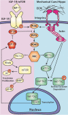
FIG. 1.
FIG. 1.
Crosstalk between the IGF-1R/mTOR and Hippo pathways via the mechanical microenvironment. The canonical IGF-1R/mTOR pathway (left; yellow and orange proteins; pink backdrop) shares substantial interaction with various mechanosensitive proteins (right; pink, orange, and green proteins; blue backdrop). Mechanical signals are generated from integrin binding and from the stress on the actin cytoskeleton (pink proteins). Several interactors are involved in both signal cascades (orange proteins). Nuclear YAP/TAZ promotes signaling through mTOR by inhibiting PTEN through microRNA (red proteins and signal molecules).
FIG. 2.
FIG. 2.
The intersection of biological and physical elements in the tumor microenvironment. The constituent elements of the tumor microenvironment can be roughly categorized by their biological or physical nature with crosstalk between these nonexclusive categories. Among biological elements (top; pink backdrop), heterotypic cell signaling through juxtacrine or paracrine signals with bioactive factors (orange) are critical to the tumor microenvironment. These cells may include stromal cells such as fibroblasts (green) or bone marrow cells (blue) and immune cells such as lymphocytes (pink) and macrophages (yellow), all of which may aid in tumor growth and the evasion of immune surveillance. Critically, vascular elements with leaky endothelial vessels (red) contribute to a nutrient gradient (light orange) in tumor niches. Leaky vessels also provide physical elements (bottom; blue backdrop) in the tumor microenvironment by contributing to oxygen gradients (magenta) and pH changes (yellow) as well as a mode of hematogenous spread. Extracellular matrix (brown) protein signaling provides both biological signals to tumor cells and the physical substrate with which tumor cells interact; therefore, signals from these tumor elements straddle the pink and blue border. The tumor architecture itself (brown) will be dictated by the tissue type as well as the extracellular matrix structures that arise as a result of the tumor response. Finally, interstitial fluid flow that inherent to the native tissue type or due to leaky vessels (blue arrows) also has been shown to contribute to pathogenic signaling in sarcoma.
FIG. 3.
FIG. 3.
Overview of tissue-engineered preclinical models of cancer. Engineered tumor models may be classified into three major groups. The least complex group consists of monolayer culture systems (left) including tissue culture plates and transwell systems. Tissue-engineered 3D models (center) include a variety of approaches to facilitate the growth of cells in three dimensions. Suspension culture with clusters of cells proliferating as spheroids or organoids was some of the first models to be developed. Tissue engineers have used scaffold fabrication (hydrogels, electrospun scaffolds, 3D printing) and/or microfluidic fabrication techniques to incorporate critical elements of the microenvironment. In vivo+ models (right) include animal models as well as decellularized tissue models that are more complex in form and structure than monolayer or 3D models but generally have lower throughput. The advantages and disadvantages of each model are discussed in Table 1.

References

    1. Prasad V., and Mailankody S.. Research and Development Spending to Bring a Single Cancer Drug to Market and Revenues After Approval. JAMA Intern Med 177, 1569, 2017 - PMC - PubMed
    1. Wong C.H., Siah K.W., and Lo A.W.. Estimation of clinical trial success rates and related parameters. Biostatistics 2018 - PMC - PubMed
    1. Hartung T. Look back in anger - what clinical studies tell us about preclinical work. ALTEX 30, 275, 2013 - PMC - PubMed
    1. Mak I.W., Evaniew N., and Ghert M.. Lost in translation: animal models and clinical trials in cancer treatment. Am J Transl Res 6, 114, 2014 - PMC - PubMed
    1. Hanahan D., and Weinberg R.A.. Hallmarks of cancer: the next generation. Cell 144, 646, 2011 - PubMed

Publication types